Review
. 2020 Jun;43(6):433-449.
doi: 10.1016/j.tins.2020.03.012. Epub 2020 Apr 17.

Microglia versus Monocytes: Distinct Roles in Degenerative Diseases of the Retina

Affiliations
Review

Microglia versus Monocytes: Distinct Roles in Degenerative Diseases of the Retina

Chen Yu et al. Trends Neurosci. 2020 Jun.

Abstract

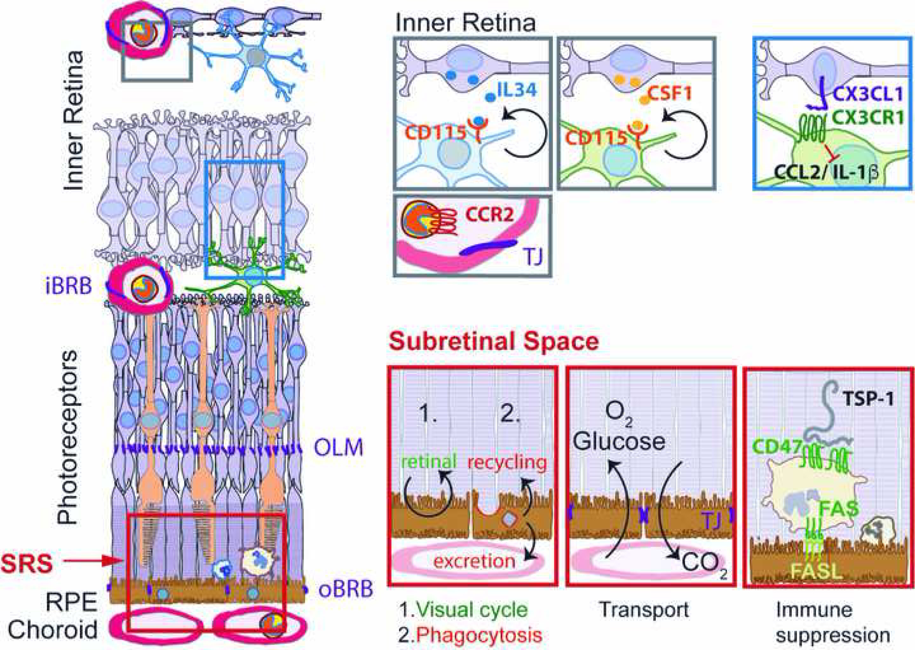
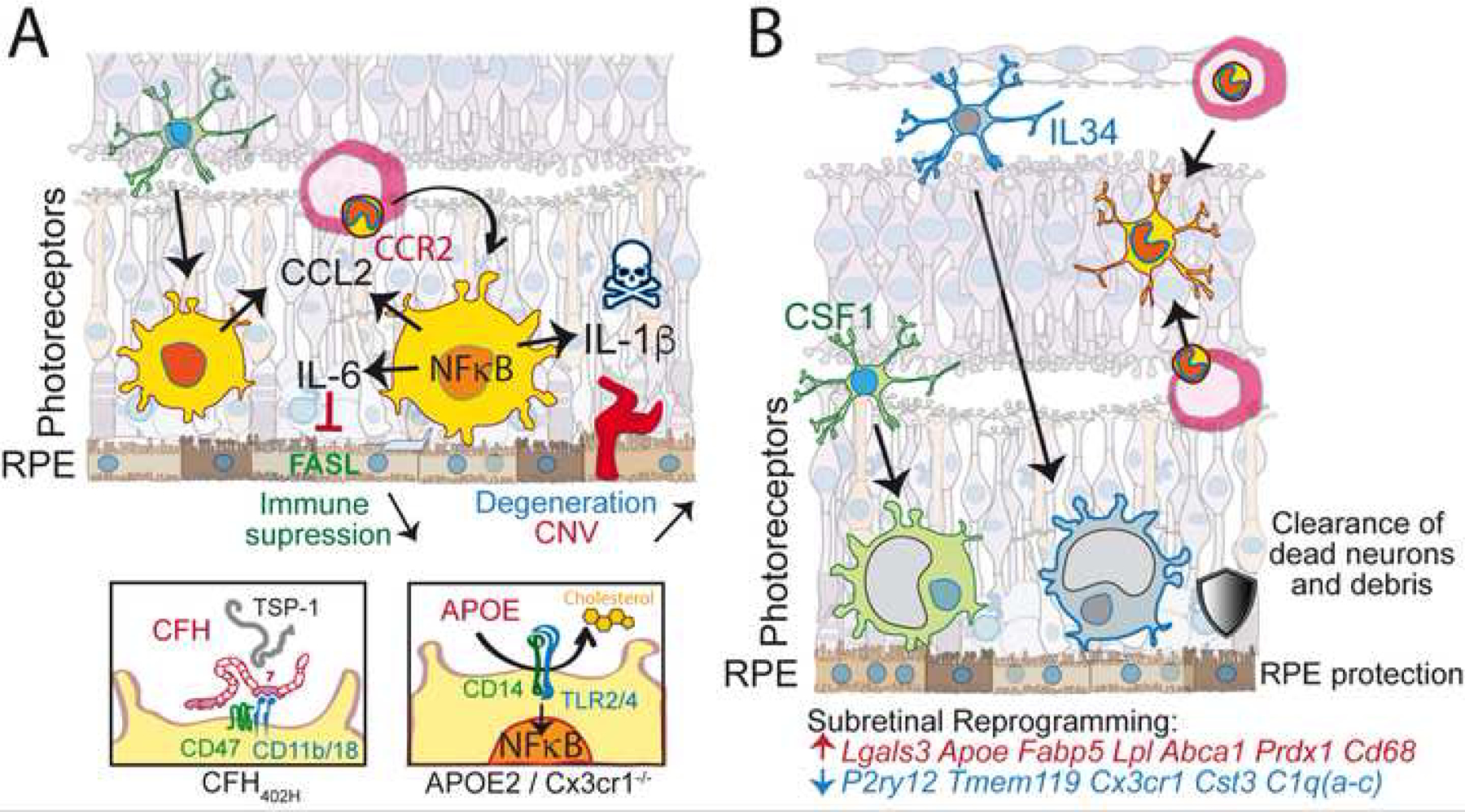
Unlike in the healthy mammalian retina, macrophages in retinal degenerative states are not solely comprised of microglia but may include monocyte-derived recruits. Recent studies have applied transgenics, lineage-tracing, and transcriptomics to help decipher the distinct roles of these two cell types in the diseasesettings of inherited retinal degenerations and age-related macular degeneration.Literature discussed here focuses on the ectopic presence of both macrophage types in the extracellular site surrounding the outer aspect ofphotoreceptor cells (i.e.,the subretinal space), which is crucially involved in the pathobiology. From these studies we propose a working model in which perturbed photoreceptor states cause microglial dominant migration to the subretinal space as a protective response, whereas the abundant presence ofmonocyte-derived cells there instead drives and accelerates pathology. The latter, we propose, is underpinned by specific genetic and nongenetic determinants that lead to a maladaptive macrophage state.

Keywords: AMD; age-related macular degeneration; inherited retinal dystrophy; macrophages; retinitis pigmentosa.


Figures

Figure 1:
Figure 1:. Physiological distribution of retinal MNPs and main functions of the subretinal space:
Physiologically the retina is only populated by IL34-dependent and CSF-1-dependent microglial cells. CCR2-positive Mos are restricted to the circulation separated from the retina by the inner and outer blood retinal barrier. CX3CL1 is constitutively expressed as a transmembrane protein in inner retinal neurons and provides a tonic inhibitory signal to the microglial checkpoint factor CX3CR1, expressed by retinal MCs. The subretinal space, demarcated by the tight junctions of the outer limiting membrane and of the RPE is the site of 1.) the visual cycle; 2.) phagocytosis of used photoreceptor outer segments and excretion or recycling the material; 3.) Oxygen and glucose uptake and CO2 excretion; 4.) immune suppression, in which FasL induces elimination of infiltrating leukocytes after activation of the integrin associated protein CD47 by thrombospondin 1 (TSP-1). iBRB: inner blood brain barrier; oBRB: outer blood brain barrier; OLM: outer limiting membrane; SRS: subretinal space; RPE: retinal pigment epithelium; IL: interleukin; CSF: colony stimulating factor; TJ; tight junction.
Figure 2:
Figure 2:. Molecular mechanisms of the role of MNPs in retinal degenerations:
(A) participation of mononuclear-derived cells in the infiltration of the SRS: loss of microglial checkpoint genes (such as in Cx3cr1 deficiency) lowers the threshold of microglial activation and induces NF-κB signaling and inflammatory cytokine secretion in conditions of normally sub-threshold stress. Increased levels of CCL2, IL-6 and IL-1β lead to monocyte recruitment, down-regulation of FasL and immune-suppression and photoreceptor degeneration and neovascularization, respectively. (A’) In AMD this NF-κB activation can be due to the AMD-associated APOE2 allele that, similar to Cx3cr1-deficiency in mice, leads to increased APOE concentrations, lipid-raft destabilization of the plasma membrane and the activation of the innate immunity receptor cluster (CD14 and TLR2/4). (A”) Furthermore, the AMD-associated Complement Factor H 402H variant inhibits MNP elimination as it blocks TSP-1 mediated CD47 activation particularly efficiently. Both AMD-risk factors thereby promote pathogenic subretinal inflammation. (B) Dominance of resident microglia in the SRS in Cx3cr1 sufficient settings: both IL34-dependent and independent microglia migrate to the SRS, which is poorly accessible to MdCs. These subretinal microglia are transcriptionally reprogrammed by downregulating homeostatic marker genes and upregulating genes involved in lipid metabolism, anti-oxidant activity and others as indicated. Functionally, these cells clear dead neurons and cellular debris and protect the RPE and photoreceptors.
Figure 3:
Figure 3:. A unified hypothesis for MNP responses in IRD and AMD:
Adaptive responses by resident microglia express low levels of pathogenic cytokines, help eliminate toxic waste and slow down degeneration. When confounded by risk factors, such as genetic variants and environmental factors, microglia more readily express pathogenic cytokines and recruit monocytes. Such maladaptive responses by MNPs exacerbate retinal degenerative diseases.

References

    1. Ginhoux F et al. (2010) Fate mapping analysis reveals that adult microglia derive from primitive macrophages. Science 330 (6005), 841–5. - PMC - PubMed
    1. Reyes NJ et al. (2017) New insights into mononuclear phagocyte biology from the visual system. Nat Rev Immunol 17 (5), 322–332. - PMC - PubMed
    1. O’Koren EG et al. (2019) Microglial Function Is Distinct in Different Anatomical Locations during Retinal Homeostasis and Degeneration. Immunity 50 (3), 723–737 e7. - PMC - PubMed
    1. Goldmann T et al. (2016) Origin, fate and dynamics of macrophages at central nervous system interfaces. Nat Immunol 17 (7), 797–805. - PMC - PubMed
    1. McMenamin PG et al. (2019) Immune cells in the retina and choroid: Two different tissue environments that require different defenses and surveillance. Prog Retin Eye Res 70, 85–98. - PMC - PubMed

Publication types