. 2020 Jun:56:102786.
doi: 10.1016/j.ebiom.2020.102786. Epub 2020 May 24.

Patient-derived pancreatic tumour organoids identify therapeutic responses to oncolytic adenoviruses

Affiliations

Patient-derived pancreatic tumour organoids identify therapeutic responses to oncolytic adenoviruses

Giulia Raimondi et al. EBioMedicine. 2020 Jun.

Abstract

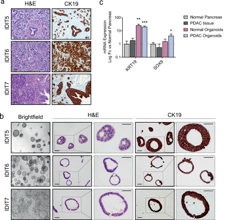
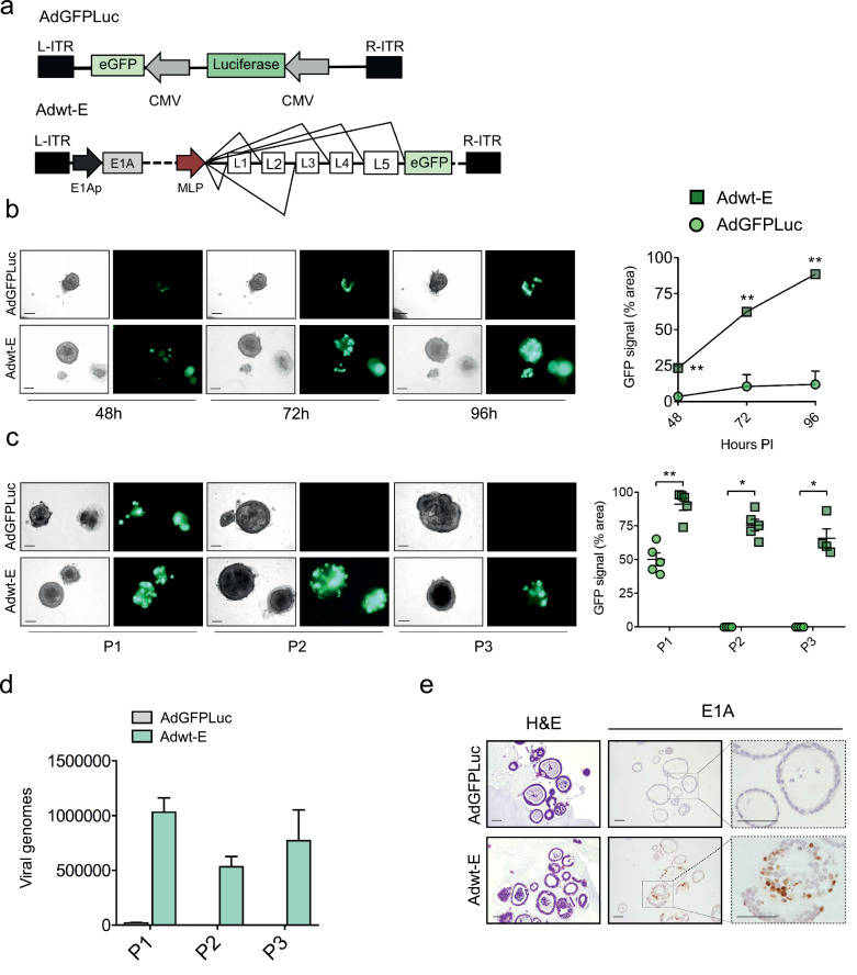
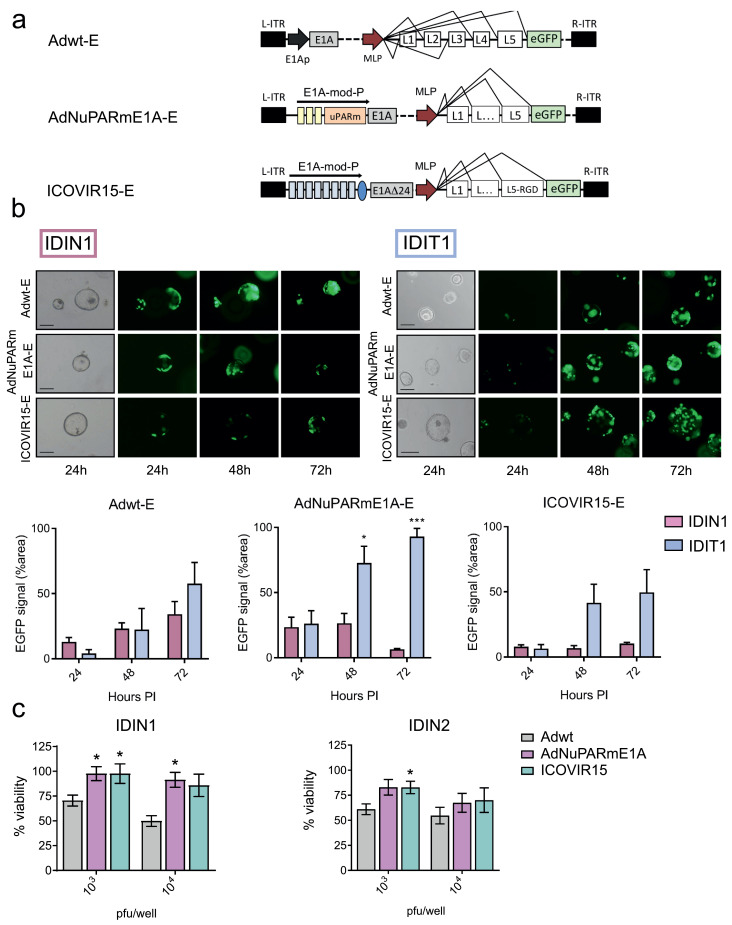
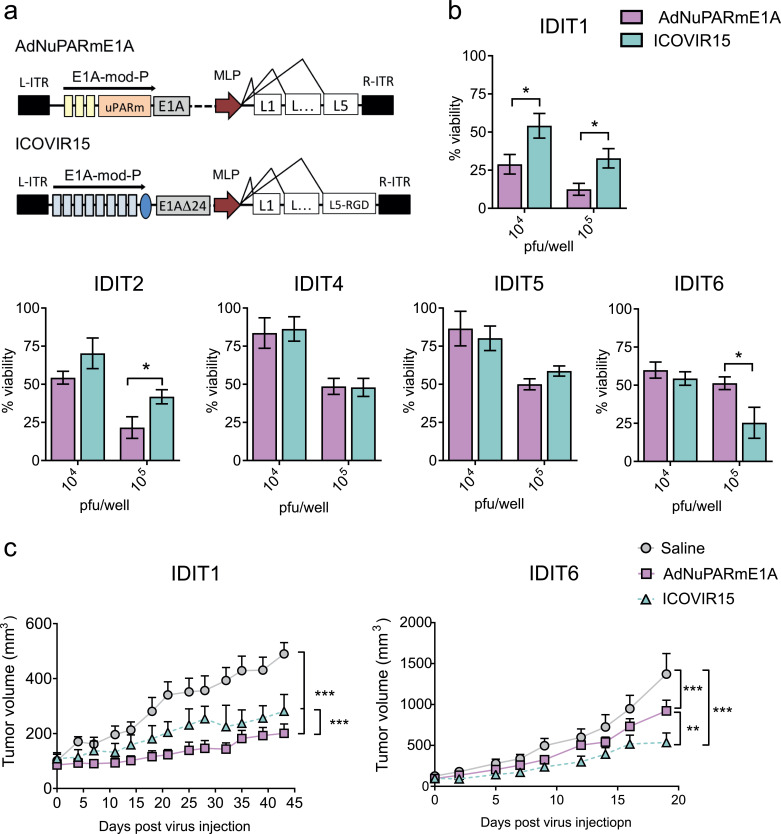
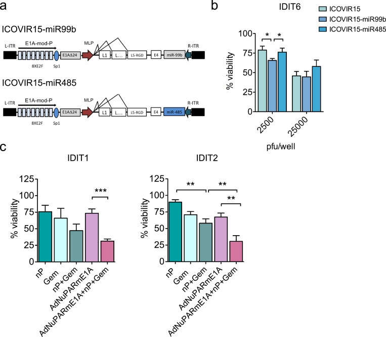
Background: Pancreatic patient-derived organoids (PDOs) are a well-established model for studying pancreatic ductal adenocarcinoma (PDAC) carcinogenesis and are potential predictors of clinical responses to chemotherapy. Oncolytic virotherapy is envisioned as a novel treatment modality for pancreatic cancer, and candidate viruses are being tested in clinical trials. Here, we explore the feasibility of using PDOs as a screening platform for the oncolytic adenovirus (OA) response.

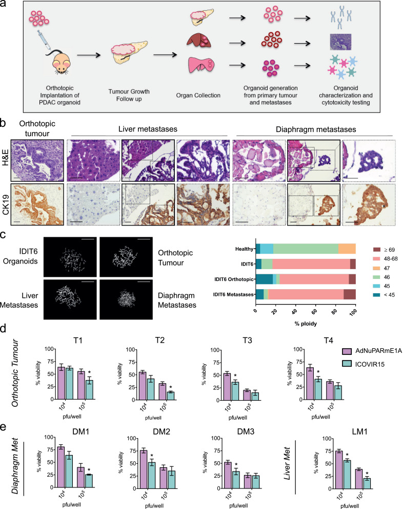
Methods: Organoids were established from healthy pancreas and PDAC tissues and assessed for infectivity, oncoselectivity, and patient-dependent sensitivity to OA. Antitumour effects were studied in vivo in organoid xenografts. Further evaluation of oncolytic responses was conducted in organoids derived from orthotopic models or metastastic tissues.

Findings: Oncolytic adenoviruses display good selectivity, with replication only in organoids derived from PDAC tumours. Furthermore, responses of PDOs to a set of OAs reveal individual differences in cytotoxicity as well as in synergism with standard chemotherapy. Adenoviral cytotoxicity in PDOs is predictive of antitumour efficacy in a subcutaneous xenograft setting. Organoids from orthotopic tumours and metastases in nude mice mirror the viral preference of PDOs, indicating that PDO sensitivity to OAs could be informative about responses in both primary tumours and metastatic foci.

Interpretation: Our data imply that pancreatic PDOs can serve as predictive tools for screening for sensitivity to OA.

Keywords: Oncolytic adenovirus (OA); Orthotopic tumours; Pancreatic ductal adenocarcinoma (PDAC); Patient-derived organoids (PDO).


Conflict of interest statement

Declaration of Competing Interest SFB and HC declare that they hold a patent (WO2015/173425). All other authors declare no conflict of interest.

Figures

Fig 1
Fig. 1
Human PDAC-derived organoids maintain characteristics of their tumour-of-origin and are enriched in ductal markers. (a) Representative histological sections of human pancreatic tumours. Left, H&E staining; right, CK19 IHC staining (scale bar 50 µm). (b) PDAC-derived organoids from matching tumours. Left, brightfield images (scale bar 50 µm); centre, H&E staining; right, CK19 IHC staining (scale bar 50 µm). (c) qRT-PCR analysis of ductal lineage markers (KRT19 and SOX9) from PDAC organoids (n = 7), normal pancreas organoids (n = 4), tumour tissue-of-origin (n = 4), and adjacent normal pancreas (n = 6). Data are represented as mean ± SEM; *p < 0.5, **p < 0.01, ***p < 0.05, non-parametric Mann–Whitney U test.
Fig 2
Fig. 2
Adenoviruses efficiently replicate in PDAC organoids. (a) Schematic representation of adenoviruses used in the experiment: the reporter non-replicative adenovirus AdGFPLuc and the replicative-competent Adwt-E, both expressing GFP. (b) Time-course GFP expression in tumour organoids infected with 5 × 105 pfu/well of AdGFPLuc or Adwt-E. Left panel shows representative images of IDIT4 organoids at the indicated time-points after infection (scale bar 100 μm). Right panel represents GFP quantification. Values are expressed as percentage of fluorescent area at each time point, normalized by total area of single organoids. Data are represented as mean ± SEM, (n > 3); **p < 0.01, ***p < 0.05 Non-parametric Mann-Whitney U Test. (c) Adenoviral propagation through passages. Organoids were infected with 5 × 105 pfu/well of AdGFPLuc or Adwt-E and 5 days later (P1) were harvested, followed by three cycles of freeze and thaw. Supernatants were used to infect fresh organoids and 5 days later (P2) the same procedure was repeated to generate P3. GFP expression was visualized at P1, P2 and P3 in organoids infected with Adwt-E, but only at P1 in AdGFPLuc infected organoids. Left panel shows representative images of organoids at P1, P2, P3 (scale bar 100 μm). Right panel represents GFP quantification at each passage. Values are expressed as percentage of fluorescent area for each time point, normalized by total area of single organoids. Data are represented as mean ± SEM, n > 3; * p < 0.05, ** p < 0.01, Non-parametric Mann-Whitney U Test. (d) Quantification of viral genomes content in P1, P2 and P3 organoid passages infected with AdGFPLuc or Adwt-E (n = 3). e. Histological sections of IDIT4 organoids infected with AdGFPLuc or Adwt-E. H&E (left panel), E1A immunostaining (right panel), scale bar 50 μm.
Fig 3
Fig. 3
Oncolytic adenoviruses selectively replicate in PDAC derived organoids. (a) Schematic representation of Adwt-E, AdNuPARmE1A-E and ICOVIR15-E adenoviruses. Yellow boxes in AdNuPARmE1A-E represent SPS sequences sensitive to NOTCH activation. Blue boxes in ICOVIR15-E correspond to E2F-responsive elements. (b) Time-course GFP expression in IDIN1 (n = 3) normal organoids and IDIT1 (n = 3) tumour organoids, infected with 5 × 103 pfu/well of either adenoviruses. (Upper panels). Representative images of GFP expression in IDIT1 and IDIN1 at 24 h, 48 h, and 72 h after infection. (Lower panels). GFP quantification of organoids cultures infected with the different viruses. Values are expressed as percentage of fluorescent area for each time point, normalized for the area of single organoids. Data are represented as mean ± SEM, n ≥ 3; * p < 0.05, **p < 0.01, Student's t-test. (c) In vitro cytotoxicity of Adwt, AdNuPARmE1A and ICOVIR15 in normal pancreas organoids (IDIN1 and IDIN2). The indicated viruses have the same backbone as described in (a) but lacked the GFP gene, as shown in Fig. 4a. IDIN1 and IDIN2 organoids derived from two different patients were infected with the indicated doses and 5 days later, cell viability was assessed by MTT assay. Data are represented as mean ± SEM, n = 5; *p < 0.05, non-parametric Mann-Whitney U Test.
Fig 4
Fig. 4
PDAC organoids display heterogeneity in sensitivity to oncolytic adenoviruses. (a) Schematic representation of the oncolytic adenoviruses. (b) In vitro cytotoxicity of AdNuPARmE1A and ICOVIR15 in a battery of PDAC organoids. Organoids were infected with the indicated doses and 5 days later, cell viability was assessed by MTT assay. Data are represented as mean ± SEM, n ≥ 4; *p < 0.05, **p < 0.01, non-parametric Mann-Whitney U test. (c) In vivo antitumour effects of oncolytic adenovirus in IDIT1 and IDIT6 subcutaneous tumours. Tumour-bearing mice were intravenously injected with 5 × 1010 vp/mice of AdNuPARmE1A, ICOVIR15 or saline solution, and tumour growth was monitored. n ≥ 6 tumours/group, **p < 0.01, ***p < 0.005, Tukey range test for multiple comparison.
Fig 5
Fig. 5
Organoids identify individual response to combined therapies or armed oncolytic adenoviruses. (a) Schematic representation of ICOVIR15-miR99b and ICOVIR15-miR485 oncolytic adenoviruses. (b) IDIT6 organoids were infected with parental ICOVIR15, ICOVIR15-miR99b, or ICOVIR15-miR485 at the indicated doses. Cytotoxicity was assessed 5 days later by MTT assay. Data are represented as mean ± SEM, n ≥ 4; *p < 0.05, non-parametric Mann–Whitney U test. (c) Combination treatments. IDIT1 and IDIT2 organoids were treated with 100 nM nab-paclitaxel and 2.5 ng/ml of gemcitabine, or AdNuPARmE1A (5000 pfu/well), or all in combination; after 5 days, cell viability was assessed by MTT assay. Data are represented as mean ± SEM; n ≥ 4, **p < 0.01, ***p < 0.005, non-parametric Mann–Whitney U test.
Fig 6
Fig. 6
Primary tumours and metastases-derived organoids from PDOs implanted in nude mice mirror PDOs sensitivity to adenoviruses. (a) Schematic representation of the experiment. Six nude mice were orthotopically implanted with 5 × 105 cells. Tumour growth was followed by palpation, and after 2–3 months, animals were euthanized and organs were collected. New organoids were generated from the orthotopic tumours and their metastases and then further analysed. (b) Histological sections obtained from primary tumours and metastases stained for H&E (left) and CK19 (right); scale bar, 50 μm. (c) Karyotyping of IDIT6 original organoids and its derived orthotopic tumour and metastases. Metaphases were induced in the different lines via colcemid treatment (0.1 μg/ml) for 24 h. A minimum of 15 metaphases per sample was counted. Left panel, representative images of the different samples. Right panel, the percentage of ploidy in each group of samples. (d, e) Orthotopic tumours and metastases derived from IDIT6 organoids from four different mice were infected at the indicated doses with AdNuPARmE1A or ICOVIR15. After five days, cytotoxicity was analysed by MTT assay. Data are represented as mean ± SEM; n ≥ 5; *p < 0.05, non-parametric Mann–Whitney U test.

Comment in

Similar articles

Cited by

References

    1. Kleeff J., Korc M., Apte M., La Vecchia C., Johnson C.D., Biankin A.V. Pancreatic cancer. Nat Rev Dis Prim. 2016;2:16022. - PubMed
    1. Siegel R.L., Miller K.D., Jemal A. Cancer statistics, 2019. CA Cancer J Clin. 2019;69:7–34. - PubMed
    1. Kersten K., Visser K.E., Miltenburg M.H., Jonkers J. Genetically engineered mouse models in oncology research and cancer medicine. EMBO Mol Med. 2017;9:137–153. - PMC - PubMed
    1. Boj S.F., Hwang C.-.I., Baker L.A., Engle D.D., Tuveson D.A., Clevers H. Model organoids provide new research opportunities for ductal pancreatic cancer. Mol Cell Oncol. 2016;3 - PMC - PubMed
    1. Boj S.F., Hwang C.-.I., Baker L.A., Chio C., II, Engle D.D., Corbo V. Organoid models of human and mouse ductal pancreatic cancer. Cell. 2015;160:324–338. - PMC - PubMed

MeSH terms

Substances