Review
. 2020 Aug;75(8):1902-1917.
doi: 10.1111/all.14421. Epub 2020 Jun 16.

Epithelial cell dysfunction, a major driver of asthma development

Affiliations
Review

Epithelial cell dysfunction, a major driver of asthma development

Irene H Heijink et al. Allergy. 2020 Aug.

Abstract

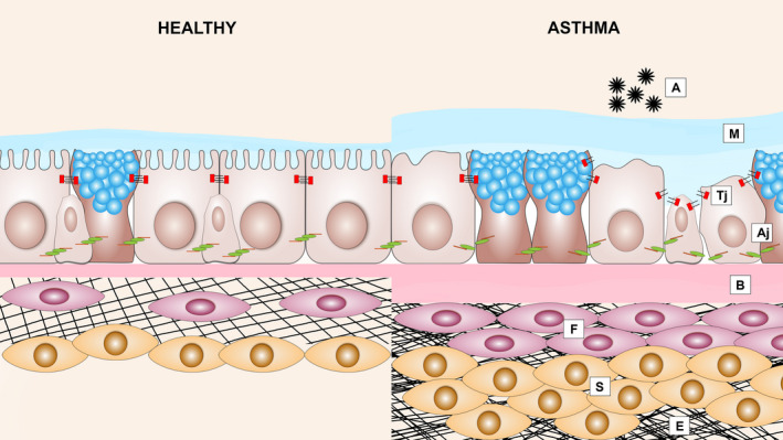
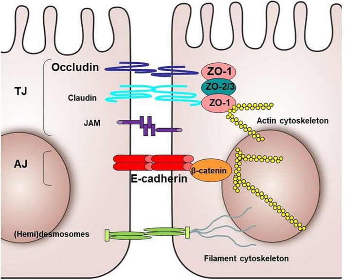
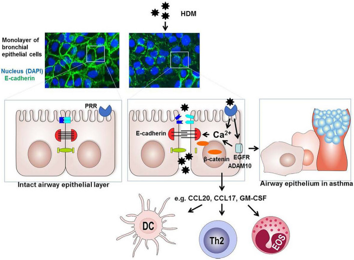
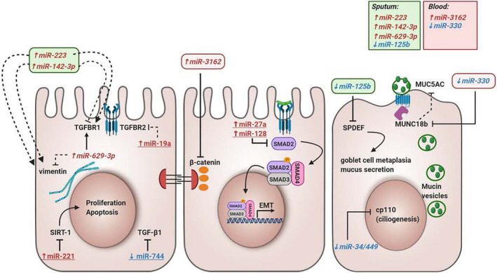
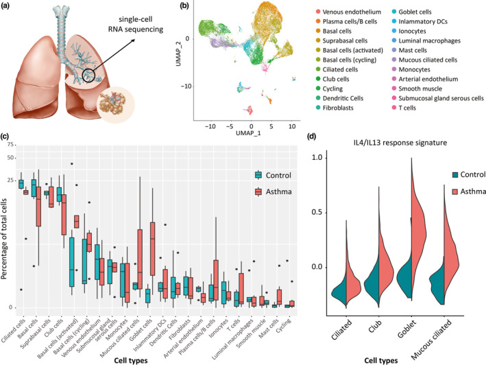
Airway epithelial barrier dysfunction is frequently observed in asthma and may have important implications. The physical barrier function of the airway epithelium is tightly interwoven with its immunomodulatory actions, while abnormal epithelial repair responses may contribute to remodelling of the airway wall. We propose that abnormalities in the airway epithelial barrier play a crucial role in the sensitization to allergens and pathogenesis of asthma. Many of the identified susceptibility genes for asthma are expressed in the airway epithelium, supporting the notion that events at the airway epithelial surface are critical for the development of the disease. However, the exact mechanisms by which the expression of epithelial susceptibility genes translates into a functionally altered response to environmental risk factors of asthma are still unknown. Interactions between genetic factors and epigenetic regulatory mechanisms may be crucial for asthma susceptibility. Understanding these mechanisms may lead to identification of novel targets for asthma intervention by targeting the airway epithelium. Moreover, exciting new insights have come from recent studies using single-cell RNA sequencing (scRNA-Seq) to study the airway epithelium in asthma. This review focuses on the role of airway epithelial barrier function in the susceptibility to develop asthma and novel insights in the modulation of epithelial cell dysfunction in asthma.

Keywords: (epi)genetics; airway remodelling; asthma; epithelial barrier; type 2 responses.


Conflict of interest statement

Dr Maes reports grants from Ghent University, Fund for Scientific Research Flanders (FWO; G053516N, G041819N, FWO‐EOS project G0G2318N), during the conduct of the study; personal fees from GlaxoSmithKline, outside the submitted work, and is shareholder of Oryzon Genomics and of Mendelion Lifesciences SL; Prof. Nawijn reports grants from the Netherlands Lung Foundation (LF 14.020 and LF 18.226), during the conduct of the study. Outside of the submitted work, Prof. Sayers laboratory reports grants from Asthma UK, British Lung Foundation Nottingham University Hospitals, National Institute for Health Research, Medical Research Council, GlaxoSmithKline and Boehringer Ingelheim; Prof. Nawijn reports grants from GSK; Prof. Heijink reports grants from the Netherlands Lung Foundation (LF 15.017) and Boehringer Ingelheim.

Figures

FIGURE 1
FIGURE 1
Structural changes in the airways of allergic asthma patients: Epithelial barrier dysfunction and airway remodelling. Asthmatic airway epithelium exposed to allergens (A) results in the disruption of adherens junctions (Aj) and tight junctions (Tj), which is accompanied by loss of ciliated cells, mucus hypersecretion (M), thickening of the basal membrane (B), subepithelial fibrosis (F), increased smooth muscle mass (S) and excessive deposition of ECM (E)
FIGURE 2
FIGURE 2
Schematic representation of the basic structural components of epithelial junctions. AJ, Adherens Junction; JAM, junctional adhesion molecule; TJ, Tight junction
FIGURE 3
FIGURE 3
Proposed model of house dust mite (HDM)‐induced airway epithelium barrier dysfunction. Allergens including HDM can directly cleave epithelial junctions proteolytically or act on various pattern recognition receptors (PRRs), including PAR‐2, C‐type lectins (CLR) and purinergic receptors. Their activation can induce degradation and/or delocalization of junctional proteins, including E‐cadherin, in which intracellular Ca2+ signalling and subsequent activation of calpain may be involved and epidermal growth factor receptor (EGFR) activation. 161 EGFR can activate ADAM10, a sheddase of E‐cadherin as well as CCL20. 40 In addition, EGFR signalling can induce secretion of pro‐inflammatory mediators, such as CCL20, CCL17 and GM‐CSF that attract and/or activate dendritic cells (DCs), Th2 cells and eosinophils (EOS). 22 When epithelial repair and re‐differentiation is impaired, persistent loss of E‐cadherin can result in activation of β‐catenin‐mediated programs that cause further loss of epithelial characteristics, induction of a more basal/mesenchymal phenotype as well as goblet cell hyperplasia, with loss of ciliated cells, as is also characteristic of the epithelial phenotype in asthma
FIGURE 4
FIGURE 4
The influence of microRNAs in epithelial barrier function. This overview illustrates miRNAs that are differentially expressed in asthma and could contribute to epithelial barrier dysfunction in asthma. miRNAs coloured in red with upward arrow are upregulated in asthma, and miRNAs coloured in blue with downward arrow are downregulated in asthma. miRNAs with an underscore were measured in bronchial epithelial cells, and miRNAs in italic were measured in sputum or blood from asthma patients and controls. Black lines ending with a perpendicular line indicate inhibitory effects, and black lines ending with an arrow indicate a stimulatory effect. Full lines indicate direct effects, and half‐full lines indicate indirect effects. EMT, epithelial‐mesenchymal transition; LPS, lipopolysaccharide; SIRT‐1, Sirtuin 1; SPDEF, SAM Pointed Domain Containing ETS Transcription Factor; TGF‐β1, Transforming Growth Factor Beta 1; TGFBR1, Transforming Growth Factor Beta Receptor 1
FIGURE 5
FIGURE 5
Analysis of airway epithelial cells in asthma using single‐cell RNA sequencing. (A) Airway wall biopsies are obtained from 5th‐7th generation airway through bronchoschopy, followed by tissue digestion and scRNA‐Seq analysis. (B) Unsupervised clustering identifies a large number of epithelial and nonepithelial cell types from airway wall. (C) Comparison of relative frequencies of cell types identified increased number of goblet cells and mucous ciliated cells, a novel, disease‐associated ciliated epithelial cell phenotype and increased numbers of mast cells and B cells in asthma compared to healthy. (D) Analysis of epithelial cell subset‐specific transcriptomes reveals presence of IL4/IL13‐induced gene transcription in goblet cells and mucous ciliated cells, specifically in asthma

References

    1. The Global Asthma Report 2018; 2018. http://www.globalasthmareport.org/. Accessed January 14, 2020.
    1. Heijink IH, Nawijn MC, Hackett TL. Airway epithelial barrier function regulates the pathogenesis of allergic asthma. Clin Exp Allergy. 2014;44(5):620‐630. - PubMed
    1. Eder W, Ege MJ, von Mutius E. The asthma epidemic. N Engl J Med. 2006;355(21):2226‐2235. - PubMed
    1. Dunican EM, Fahy JV. The role of type 2 inflammation in the pathogenesis of asthma exacerbations. Ann Am Thorac Soc. 2015;12(Suppl 2):S144‐S149. - PMC - PubMed
    1. Svenningsen S, Nair P. Asthma endotypes and an overview of targeted therapy for asthma. Front Med (Lausanne). 2017;4:158. - PMC - PubMed