Modeling the natural history of ductal carcinoma in situ based on population data
- PMID: 32460821
- PMCID: PMC7251719
- DOI: 10.1186/s13058-020-01287-6
Modeling the natural history of ductal carcinoma in situ based on population data
Abstract
Background: The incidence of ductal carcinoma in situ (DCIS) has increased substantially since the introduction of mammography screening. Nevertheless, little is known about the natural history of preclinical DCIS in the absence of biopsy or complete excision.
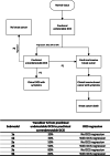
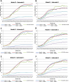
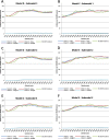
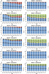
Methods: Two well-established population models evaluated six possible DCIS natural history submodels. The submodels assumed 30%, 50%, or 80% of breast lesions progress from undetectable DCIS to preclinical screen-detectable DCIS; each model additionally allowed or prohibited DCIS regression. Preclinical screen-detectable DCIS could also progress to clinical DCIS or invasive breast cancer (IBC). Applying US population screening dissemination patterns, the models projected age-specific DCIS and IBC incidence that were compared to Surveillance, Epidemiology, and End Results data. Models estimated mean sojourn time (MST) in the preclinical screen-detectable DCIS state, overdiagnosis, and the risk of progression from preclinical screen-detectable DCIS.
Results: Without biopsy and surgical excision, the majority of DCIS (64-100%) in the preclinical screen-detectable state progressed to IBC in submodels assuming no DCIS regression (36-100% in submodels allowing for DCIS regression). DCIS overdiagnosis differed substantially between models and submodels, 3.1-65.8%. IBC overdiagnosis ranged 1.3-2.4%. Submodels assuming DCIS regression resulted in a higher DCIS overdiagnosis than submodels without DCIS regression. MST for progressive DCIS varied between 0.2 and 2.5 years.
Conclusions: Our findings suggest that the majority of screen-detectable but unbiopsied preclinical DCIS lesions progress to IBC and that the MST is relatively short. Nevertheless, due to the heterogeneity of DCIS, more research is needed to understand the progression of DCIS by grades and molecular subtypes.
Keywords: Breast carcinoma in situ; Breast neoplasms; Disease progression; Early detection of cancer; United States.
Conflict of interest statement
The authors declare that they have no competing interests.
Figures


References
-
- Oseni TO. Zhang B, Coopey SB, Gadd MA, Hughes KS, Chang DC. Twenty-five year trends in the incidence of ductal carcinoma in situ in US women. J Am Coll Surg. 2019;228(6):932–939. - PubMed
-
- Maxwell AJ, Clements MK, Hilton MB, Dodwell DJ, Evans A, Kearins MO, et al. Risk factors for the development of invasive cancer in unresected ductal carcinoma in situ. Eur J Surg Oncol. 2018;44(4):429–435. - PubMed
-
- Erbas B, Provenzano E, Armes J, Gertig D. The natural history of ductal carcinoma in situ of the breast: a review. Breast Cancer Res Treat. 2006;97(2):135–144. - PubMed
-
- Morrow M, Strom EA, Bassett LW, Dershaw DD, Fowble B, Harris JR, et al. Standard for the management of ductal carcinoma in situ of the breast (DCIS) CA Cancer J Clin. 2002;52(5):256–276. - PubMed
Publication types
MeSH terms
Grants and funding
LinkOut - more resources
Full Text Sources
Medical

