. 2020 May 27;11(1):206.
doi: 10.1186/s13287-020-01719-2.

Three-dimensional culture of MSCs produces exosomes with improved yield and enhanced therapeutic efficacy for cisplatin-induced acute kidney injury

Affiliations

Three-dimensional culture of MSCs produces exosomes with improved yield and enhanced therapeutic efficacy for cisplatin-induced acute kidney injury

Jingyuan Cao et al. Stem Cell Res Ther. .

Abstract

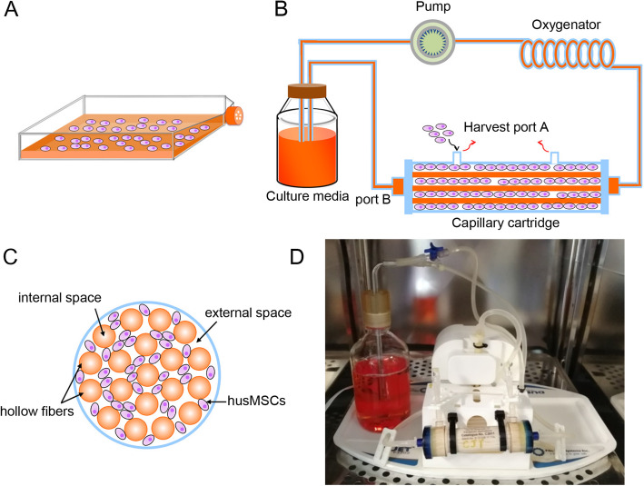
Background: Exosomes derived from mesenchymal stem cells (MSC-exos) have been demonstrated with great potential in the treatment of multiple human diseases including acute kidney injury (AKI) by virtue of their intrinsic cargoes. However, there are major challenges of low yield and the lack of an established biomanufacturing platform to efficiently produce MSC-exos, thereby limiting their therapeutic application. Here, we aimed to establish a novel strategy to produce MSC-exos with a hollow fiber bioreactor-based three-dimensional (3D) culture system and evaluate the therapeutic efficacy of 3D-exosomes (3D-exos) on AKI.

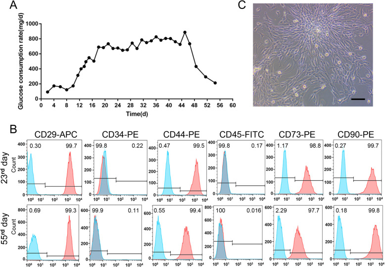
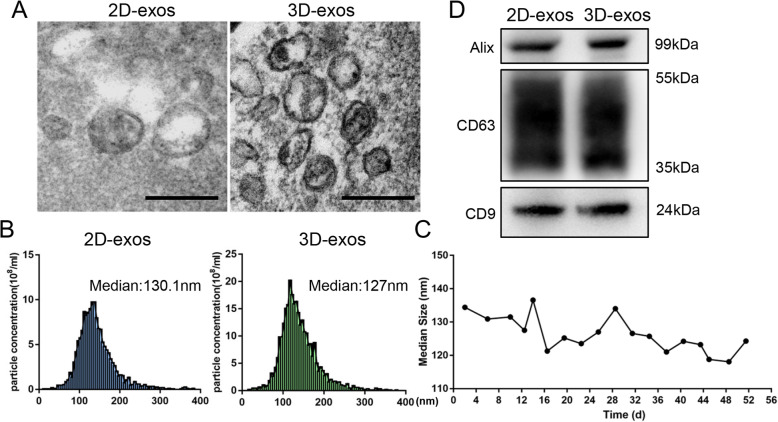
Methods: Mesenchymal stem cells (MSCs) were isolated from fresh human umbilical cord and cultured in two-dimensional (2D) flasks. 2 × 10⁸ MSCs were inoculated into the hollow fiber bioreactor for 3D culture. The culture supernatants were collected every 1 or 2 days for isolating exosomes. Exosomes from 2D (2D-exos) and 3D cultures were characterized by transmission electron microscopy, nanoparticle tracking analysis, and western blotting analysis of exosome markers. The yield of exosomes from 2 × 10⁸ MSCs seeded in 2D and 3D culture system was compared, based on protein quantification. The therapeutic efficacy of 2D-exos and 3D-exos was investigated in a murine model of cisplatin-induced AKI in vivo and in vitro.

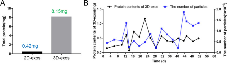
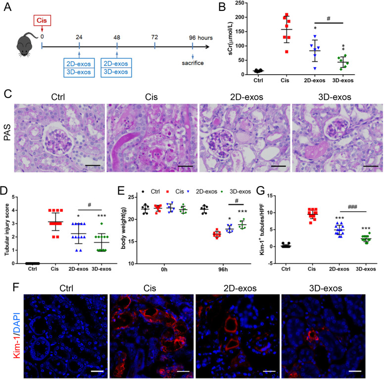
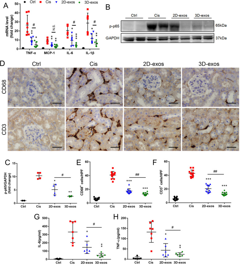
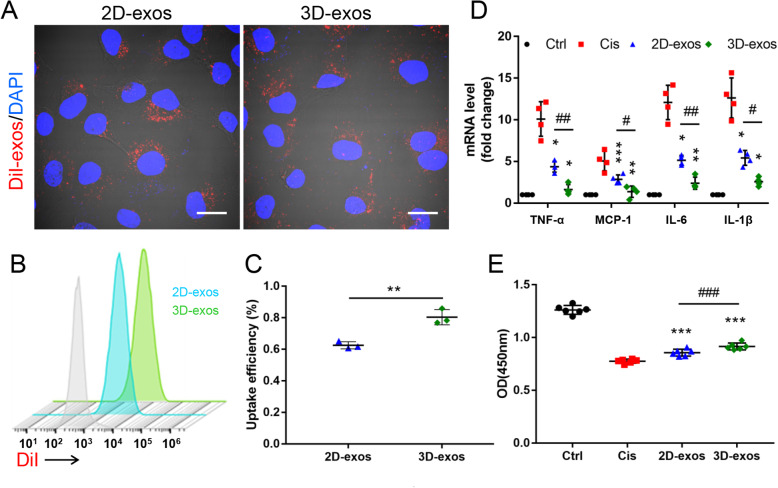
Results: 3D culture did not significantly change the surface markers of MSCs, as well as the morphology, size, and exosomal markers of 3D-exos when compared to those of 2D-exos. Compared with conventional 2D culture, the 3D culture system increased total exosome production up to 19.4-fold. 3D-exos were more concentrated in the harvested supernatants (15.5-fold) than 2D-exos, which led to a higher exosome collection efficiency of 3D culture system. In vivo, both 2D-exos and 3D-exos significantly alleviated cisplatin-induced murine AKI evidenced by improved renal function, attenuated pathological changes of renal tubules, reduced inflammatory factors, and repressed T cell and macrophage infiltration. Impressively, 3D-exos were more effective than 2D-exos. Moreover, 3D-exos were taken up by tubular epithelial cells (TECs) with improved efficiency, thereby exhibiting superior anti-inflammatory effect and improved viability of TECs in vitro.

Conclusions: In summary, our findings demonstrate that the hollow fiber 3D culture system provides an efficient strategy for the continuous production of MSC-exos which has enhanced therapeutic potential for cisplatin-induced AKI.

Keywords: Acute kidney injury; Exosomes; Mesenchymal stem cell; Three-dimensional culture.


Conflict of interest statement

The authors declare that they have no competing interests.

Figures

Fig. 1
Fig. 1
Schematic and photograph of the 2D and 3D culture system. a The schematic of conventional 2D flask. b The general schematic of the hollow fiber bioreactor-based 3D culture system. The system was composed of a pulsatile perfusion pump, an oxygenator, a cartridge containing thousands of hollow fibers, a bottle of culture medium, and the connecting tube. c A general schematic of cross-sectional view through the bioreactor. d The photograph of our 3D culture system
Fig. 2
Fig. 2
3D Culture of hucMSCs. a The glucose consumption rate of 3D culture system. b The flow cytometry analysis of MSC (CD29, CD44, CD73, and CD90) and HSC (CD34 and CD45) surface markers of exfoliated cells on the 23rd and 55th days. Blue solid peaks represent the isotype controls, and the red solid peaks represent the marker indicated. c The optical micrograph of exfoliated cells in the supernatants from 3D system. Scale bar, 50 μm
Fig. 3
Fig. 3
Characterization of 2D-exos and 3D-exos. a Morphology of 2D-exos and 3D-exos under TEM. Scale bar, 200 nm. b Representative NTA analysis of the particle size distribution of 2D-exos and 3D-exos. The difference between the median diameter of 2D-exos (n = 4) and 3D-exos (n = 18) was not statistically significant (p > 0.05). c The particle size distribution of 3D-exos was detected at different time points, and the median diameters of 3D-exos were 118.1–141.3 nm. d Western blotting analysis of exosomal surface markers (CD9, CD63, and Alix) of 2D-exos and 3D-exos
Fig. 4
Fig. 4
The exosomes yield of 2D-culture and 3D-culture. a The total output of exosomes from 2 × 108 MSCs seeded in 2D and 3D culture system. b The number of particles and protein contents of 3D-exos harvested from 3D culture system at different time points
Fig. 5
Fig. 5
The therapeutic efficacy of 2D-exos and 3D-exos in cisplatin-induced AKI mice model. a Schematic diagram of the experimental design. In brief, mice were concurrently treated with PBS, 2D-exos (100 μg) or 3D-exos (100 μg) at 24 h and 48 h after cisplatin injection, and were sacrificed at 96 h after disease induction. b Effects of 2D-exos and 3D-exos on serum creatinine (n = 6–7). c Representative images of PAS staining of renal cortex. Scale bar, 50 μm. d The quantification of tubular injury based on PAS staining (n = 6–7). e Effects of 2D-exos and 3D-exos on the body weight (n = 6–7). f Representative confocal images of kidney injury molecular-1(Kim-1) in tubules. Scale bar, 25 μm. g The quantification of Kim-1+ tubules per HPF (n = 6). Data are presented as mean ± SD, *p < 0.05, **p < 0.01, ***p < 0.001 vs. Cis group, #p < 0.05, ###p < 0.001
Fig. 6
Fig. 6
Enhanced anti-inflammatory efficacy of 3D-exos in cisplatin-treated mice. a Real-time PCR analysis of inflammatory cytokine mRNA levels in kidney tissues (n = 6–7). b Western blotting analysis of p-p65 in kidney tissues. c Quantification of p-p65 in kidney tissues (n = 4). d Representative immunostaining images of CD68+ macrophages or CD3+ T cells in the tubulointerstitium. Scale bar, 50 μm. e Quantification of CD68+ macrophages in the tubulointerstitium (n = 6). f Quantification of CD3+ T cells in the tubulointerstitium (n = 6). g Levels of IL-6 in the mouse serum was detected by ELISA (n = 6–7). h Levels of TNF-α in the mouse serum was detected by ELISA (n = 6–7). Data are presented as mean ± SD, *p < 0.05, **p < 0.01, ***p < 0.001 vs. Cis group, #p < 0.05, ##p < 0.01
Fig. 7
Fig. 7
Enhanced therapeutic efficacy of 3D-exos in cisplatin-treated TECs. a TECs took up 2D-exos and 3D-exos. DiI-positive (red) cells were observed under confocal microscope. Scale bar, 10 μm. b DiI-positive cells detecting by flow cytometry. Blue solid peak represents cisplatin-treated TECs internalizing DiI-labeled 2D-exos, and the green solid peak represents cisplatin-treated TECs internalizing DiI-labeled 3D-exos. c The uptake efficiency of 2D-exos and 3D-exos. d Real-time PCR analysis of inflammatory cytokine mRNA levels in TECs (n = 4). e CCK-8 assay in TECs (n = 6). Data are presented as mean ± SD, *p < 0.05, **p < 0.01, ***p < 0.001, vs. Cis group, #p < 0.05, ##p < 0.01, ###p < 0.001

References

    1. Harrell Carl, Fellabaum Crissy, Jovicic Nemanja, Djonov Valentin, Arsenijevic Nebojsa, Volarevic Vladislav. Molecular Mechanisms Responsible for Therapeutic Potential of Mesenchymal Stem Cell-Derived Secretome. Cells. 2019;8(5):467. - PMC - PubMed
    1. Gazdic M, Volarevic V, Arsenijevic N, et al. Mesenchymal stem cells: a friend or foe in immune-mediated diseases. Stem Cell Rev Rep. 2015;11(2):280–287. - PubMed
    1. Shi Y, Wang Y, Li Q, et al. Immunoregulatory mechanisms of mesenchymal stem and stromal cells in inflammatory diseases. Nat Rev Nephrol. 2018;14(8):493–507. - PubMed
    1. Lindsay JO, Allez M, Clark M, et al. Autologous stem-cell transplantation in treatment-refractory Crohn’s disease: an analysis of pooled data from the ASTIC trial. Lancet Gastroenterol Hepatol. 2017;2(6):399–406. - PubMed
    1. Yuan X, Qin X, Wang D, et al. Mesenchymal stem cell therapy induces FLT3L and CD1c(+) dendritic cells in systemic lupus erythematosus patients. Nat Commun. 2019;10(1):2498. - PMC - PubMed

Publication types

LinkOut - more resources