Review
. 2020 Aug;98(2):314-322.
doi: 10.1016/j.kint.2020.05.013. Epub 2020 May 24.

The case of complement activation in COVID-19 multiorgan impact

Affiliations
Review

The case of complement activation in COVID-19 multiorgan impact

Marina Noris et al. Kidney Int. 2020 Aug.

Abstract

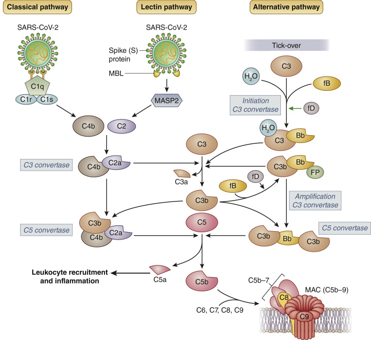
The novel coronavirus disease COVID-19 originates in the lungs, but it may extend to other organs, causing, in severe cases, multiorgan damage, including cardiac injury and acute kidney injury. In severe cases, the presence of kidney injury is associated with increased risk of death, highlighting the relevance of this organ as a target of SARS-CoV-2 infection. COVID-19-associated tissue injury is not primarily mediated by viral infection, but rather is a result of the inflammatory host immune response, which drives hypercytokinemia and aggressive inflammation that affect lung parenchymal cells, diminishing oxygen uptake, but also endothelial cells, resulting in endotheliitis and thrombotic events and intravascular coagulation. The complement system represents the first response of the host immune system to SARS-CoV-2 infection, but there is growing evidence that unrestrained activation of complement induced by the virus in the lungs and other organs plays a major role in acute and chronic inflammation, endothelial cell dysfunction, thrombus formation, and intravascular coagulation, and ultimately contributes to multiple organ failure and death. In this review, we discuss the relative role of the different complement activation products in the pathogenesis of COVID-19-associated tissue inflammation and thrombosis and propose the hypothesis that blockade of the terminal complement pathway may represent a potential therapeutic option for the prevention and treatment of lung and multiorgan damage.

Keywords: C5 inhibition; COVID-19; complement activation; complement terminal pathway; kidney injury; vascular injury.


Figures

Figure 1
Figure 1
Coronavirus disease 2019 (COVID-19) and the kidney. Severe acute respiratory syndrome coronavirus 2 (SARS-CoV-2) initially infects the lungs, where it replicates and causes cytolysis of alveolar cells and an inflammatory burst, diminishing oxygen uptake, but the virus can injure many other organs. Kidney dysfunction is common in patients with severe forms of COVID-19 and is an important risk factor for death. SARS-CoV-2 may bind directly to angiotensin-converting enzyme 2 (ACE2) expressed on kidney cells, cause cell injury, and activate the inflammatory response and the complement cascade locally (green box). Kidney damage and dysfunction may also derive from drug nephrotoxicity or systemic events like poor blood oxygenation, lowered blood pressure, or cytokine storms (orange box). AKI, acute kidney injury.
Figure 2
Figure 2
The 3 complement pathways and severe acute respiratory syndrome coronavirus 2 (SARS-CoV-2). The classical pathway is activated by the binding of natural IgM or induced IgG antibodies, which form immune complexes with viral antigens, to the complement component (C) 1 complex, comprising C1q, C1r, and C1s molecules. The lectin pathway is triggered by the binding of mannose-binding lectin (MBL) with SARS-CoV spike (S) protein, which leads to the activation of MBL-associated serine protease 2 (MASP-2). The classical and lectin pathways converge into the cleavage of C2 and C4, leading to the formation of the C3 convertase (C4bC2a) of the classical/lectin pathways. The alternative pathway is continuously activated in plasma by low-grade hydrolysis (tick-over) of C3 that forms C3(H2O). The latter binds to factor B (fB), which, in turn, is cleaved by factor D (fD) to form the alternative pathway fluid-phase initiation C3 convertase. The C3 convertases cleave C3 into C3a, an anaphylotoxin, and C3b, which deposits on cell surfaces. C3b produced by any of the 3 pathways contributes to the formation of the alternative pathway amplification C3 convertase, which cleaves additional C3 molecules, resulting in an amplification loop. In addition, C3b contributes to the formation of the C5 convertases that cleave C5, producing the anaphylatoxin C5a that attracts and activates inflammatory leukocytes, and C5b. C5b initiates the late events of complement activation, leading to the formation of the membrane-attack complex (MAC or C5b-9 complex). Bb, bb fragment of factor B; FP, properdin.
Figure 3
Figure 3
Complement activation and vasculopathy in coronavirus disease 2019 (COVID-19). Severe acute respiratory syndrome coronavirus 2 (SARS-CoV-2) binds to angiotensin-converting enzyme 2 (ACE2) on endothelial cells and activates the complement lectin pathway first and then the classical pathway, leading to complement (C) 3b deposition. C3b formation fuels the alternative pathway and participates in the formation of the C5 convertases that cleave C5 into the terminal complement products C5a and C5b-9 (upper box). The terminal complement components promote vascular inflammation through multiple processes: the anaphylatoxin C5a recruits neutrophils (polymorphonuclear neutrophils [PMN]) and monocyte/macrophages. C5b-9 and C5a induce the release of interleukin (IL)-8 and monocyte chemoattractant protein (MCP-1) by endothelial cells and stimulates the expression of adhesion molecules (ICAM-1, E-selectin, VCAM-1) that favor recruitment, adhesion, and transendothelial migration of neutrophils and macrophages. Thereafter, infiltrating leukocytes release proteases, cytokines, and reactive oxygen species (ROS) that contribute to inflammation, subendothelial matrix disruption, and remodeling, causing vasculitis-like lesions. C5a and the membrane attack complex (MAC) also induce the exocytosis of P-selectin and von Willebrand factor (VWF) multimers from endothelial cells that promote platelet adhesion, and the shedding of thrombomodulin (TM) from the endothelial cell surface, which triggers the coagulation cascade. Finally, C5b-9 directly activates platelets, causing platelet aggregation and the release of procoagulant microparticles (PMP). The results of all these events are vascular injury and dysfunction, with extensive formation of blood clots.

References

    1. Wadman M., Couzin-Frankel J., Kaiser J., Matacic C. A rampage through the body. Science. 2020;368:356–360. pages 356, 358. - PubMed
    1. Remuzzi A., Remuzzi G. COVID-19 and Italy: what next? Lancet. 2020;395:1225–1228. - PMC - PubMed
    1. Guan W.J., Ni Z.Y., Hu Y. Clinical characteristics of coronavirus disease 2019 in China. N Engl J Med. 2020;382:1708–1720. - PMC - PubMed
    1. Huang C., Wang Y., Li X. Clinical features of patients infected with 2019 novel coronavirus in Wuhan, China. Lancet. 2020;395:497–506. - PMC - PubMed
    1. Cheng Y., Luo R., Wang K. Kidney disease is associated with in-hospital death of patients with COVID-19. Kidney Int. 2020;97:829–838. - PMC - PubMed

Publication types

LinkOut - more resources