. 2020 May 27;29(156):190168.
doi: 10.1183/16000617.0168-2019. Print 2020 Jun 30.

Pulmonary capillary haemangiomatosis: a distinct entity?

Affiliations

Pulmonary capillary haemangiomatosis: a distinct entity?

Jason Weatherald et al. Eur Respir Rev. .

Abstract

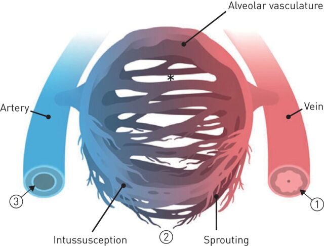
Pulmonary capillary haemangiomatosis (PCH) is a rare and incompletely understood histopathological finding characterised by abnormal capillary proliferation within the alveolar interstitium, which has long been noted to share many overlapping features with pulmonary veno-occlusive disease (PVOD). But are PCH and PVOD distinct entities that occur in isolation, or are they closely intertwined manifestations along a spectrum of the same disease? The classic clinical features of both PCH and PVOD include signs and symptoms related to pulmonary hypertension, hypoxaemia, markedly impaired diffusion capacity of the lung and abnormal chest imaging with ground glass opacities, septal lines and lymphadenopathy. In recent years, increasing evidence suggests that the clinical presentation, histopathological features, genetic substrate and pathobiological mechanisms of PCH and PVOD are overlapping and usually indistinguishable. The discovery of biallelic mutations in the eukaryotic translation initiation factor 2 α kinase 4 (EIF2AK4) gene in heritable PCH and PVOD greatly advanced our understanding of the overlapping nature of these conditions. Furthermore, recognition of PCH and PVOD-like changes in other pulmonary vascular diseases and in conditions that cause chronic pulmonary venous hyper-perfusion or hypertension suggests that PCH/PVOD may develop as a reactive process to various insults or injuries to the pulmonary vasculature, rather than being primary angiogenic disorders.


Conflict of interest statement

Conflict of interest: J. Weatherald reports grants, personal fees and non-financial support from Janssen Inc. and Actelion, personal fees and non-financial support from Bayer, personal fees from Novartis, and grants from Alberta Lung Association Canadian Vascular Network, European Respiratory Society and Canadian Thoracic Society, outside the submitted work. Conflict of interest: P. Dorfmüller has nothing to disclose. Conflict of interest: F. Perros has nothing to disclose. Conflict of interest: M-R. Ghigna has nothing to disclose. Conflict of interest: B. Girerd has nothing to disclose. Conflict of interest: M. Humbert reports personal fees and non-financial support from Acceleron, grants and personal fees from Bayer and GSK, and personal fees from Actelion, Merck and United Therapeutics, outside the submitted work. Conflict of interest: D. Montani reports grants and personal fees from Actelion and Bayer, and personal fees from GSK Pfizer, MSD and Chiesi, outside the submitted work.

Figures

FIGURE 1
FIGURE 1
a, b) Capillary haemangiomatosis foci are readily identified within lung parenchyma (star). c) They are characterised by the increase of the cell density, close to remodelled vessels. d) Alveolar septa are thickened with dilated and/or multiplication of capillaries (arrows). Increased cellularity is characterised by affluence of inflammatory cells within the alveolar septa. Endoalveolar collections of hemosiderin-laden macrophages are also visible (asterisk). Scale bars: a) 500 µm; b) 200 µm; c) 200 µm; d) 100 µm.
FIGURE 2
FIGURE 2
Pulmonary veno-occlusive disease. a) Small pulmonary veins are markedly remodelled, showing fibro-oedematous increase of the intima, leading to significant narrowing of the vascular lumen. b) Alveolar septa appear thickened with increase of the cellularity and congestion. Scale bars: 200 µm.
FIGURE 3
FIGURE 3
Prevalence of biallelic eukaryotic translation initiation factor 2 α kinase 4 (EIF2AK4) mutations in patients with familial pulmonary capillary haemangiomatosis (PCH)/pulmonary veno-occlusive disease (PVOD) and sporadic PCH/PVOD in the French Pulmonary Hypertension Network Registry. #: from 14 different families.
FIGURE 4
FIGURE 4
Interstitial remodelling patterns in pulmonary veno-occlusive disease (PVOD) eukaryotic translation initiation factor 2 α kinase 4 (EIF2AK4) mutation carriers and non-carriers. a) EIF2AK4 mutation carrier with patchy pulmonary capillary haemangiomatosis (PCH)-like foci (inset) (haematoxylin–eosin staining). b) EIF2AK4 mutation non-carrier with more diffuse PCH-like remodelling (inset) (haematoxylin–eosin staining). Scale bars=1000 µm. c) EIF2AK4 mutation carriers display a significantly higher degree of patchy PCH-like foci (p=0.014), whereas non-carriers show instead more diffuse PCH-like remodelling. EIF2AK4-C: PVOD EIF2AK4 mutation carriers; EIF2AK4-NC: PVOD EIF2AK4 non-carriers. *: p<0.05. Reproduced from [28] with permission.
FIGURE 5
FIGURE 5
Structure and architecture of pulmonary veno-occlusive disease (PVOD) and pulmonary capillary haemangiomatosis (PCH). Prominent and back-to-back proliferation of capillaries in an area with PCH a) by scanning electron microscope and b) in a complementary haematoxylin–eosin stain. c, d) Scanning electron microscope following microvascular corrosion casting of affected lung tissue reveals capillary neo-formation by sprouting (green arrow in c) and intussusceptive vascular pillars (red arrow in c) next to non-remodelled pulmonary capillaries (red frames in d). Scale bars: a) 200 µm; b) 100 µm; c) 100 µm; d) 20 µm. Reproduced from [67] with permission.
FIGURE 6
FIGURE 6
Pathogenesis of vascular remodelling in pulmonary veno-occlusive disease (PVOD). 1) Marked fibrosis of the intima and hypertrophy of the media lead to occlusion of the pulmonary postcapillary vasculature. 2) Venous occlusions cause increasing blood pressure in the pulmonary capillaries (*). Pressure overload induces excessive neoangiogenesis by sprouting and intussusceptive pillar formation likely driven by increased flow and shear stress, which results in the formation of pulmonary capillary haemangiomatosis (PCH). 3) Congestion of the pulmonary capillary and postcapillary vasculature result in pulmonary hypertension associated with subsequent sclerotic remodelling of pulmonary arteries and arterioles. Reproduced from [67] with permission.
FIGURE 7
FIGURE 7
High-resolution computed tomography scan of the chest in patients with pulmonary capillary haemangiomatosis and/or pulmonary veno-occlusive disease. a) Ground-glass opacities with centrilobular pattern, poorly defined nodular opacities, and septal lines in an adult carrier of eukaryotic translation initiation factor 2 α kinase 4 (EIF2AK4) bi-allelic mutations. b) Mediastinal lymph node enlargement in an adult carrier of EIF2AK4 bi-allelic mutations. c) A paediatric case carrying EIF2AK4 bi-allelic mutations. d) A paediatric case not carrying an EIF2AK4 bi-allelic mutation. e and f) The chest of an EIF2AK4 mutation carrier before initiation of specific therapy for pulmonary arterial hypertension showed mild abnormalities with septal lines and ground-glass opacities (e); high resolution tomography of the chest was performed 2 months after initiation of endothelin receptor antagonists for rapid worsening dyspnoea and showed a substantial increase of radiological abnormalities evocative of pulmonary oedema (f). Reproduced from [35] with permission.

Comment in

References

    1. Simonneau G, Montani D, Celermajer DS, et al. . Haemodynamic definitions and updated clinical classification of pulmonary hypertension. Eur Respir J 2019; 53: 1801913. doi:10.1183/13993003.01913-2018 - DOI - PMC - PubMed
    1. Wagenvoort CA, Beetstra A, Spijker J. Capillary haemangiomatosis of the lungs. Histopathology 1978; 2: 401–406. doi:10.1111/j.1365-2559.1978.tb01734.x - DOI - PubMed
    1. Montani D, Lau EM, Dorfmüller P, et al. . Pulmonary veno-occlusive disease. Eur Respir J 2016; 47: 1518–1534. doi:10.1183/13993003.00026-2016 - DOI - PubMed
    1. Pietra GG, Edwards WD, Kay JM, et al. . Histopathology of primary pulmonary hypertension. A qualitative and quantitative study of pulmonary blood vessels from 58 patients in the National Heart, Lung, and Blood Institute, Primary Pulmonary Hypertension Registry. Circulation 1989; 80: 1198–1206. doi:10.1161/01.CIR.80.5.1198 - DOI - PubMed
    1. Stovin PG, Mitchinson MJ. Pulmonary hypertension due to obstruction of the intrapulmonary veins. Thorax 1965; 20: 106–113. doi:10.1136/thx.20.2.106 - DOI - PMC - PubMed

MeSH terms