. 2020 Jun 3;13(6):dmm042390.
doi: 10.1242/dmm.042390.

Frameshift mutations of YPEL3 alter the sensory circuit function in Drosophila

Affiliations

Frameshift mutations of YPEL3 alter the sensory circuit function in Drosophila

Jung Hwan Kim et al. Dis Model Mech. .

Abstract

A frameshift mutation in Yippee-like (YPEL) 3 was recently found from a rare human disorder with peripheral neurological conditions including hypotonia and areflexia. The YPEL gene family is highly conserved from yeast to human, but its members' functions are poorly defined. Moreover, the pathogenicity of the human YPEL3 variant is completely unknown. We generated a Drosophila model of human YPEL3 variant and a genetic null allele of Drosophila homolog of YPEL3 (referred to as dYPEL3). Gene-trap analysis suggests that dYPEL3 is predominantly expressed in subsets of neurons, including larval nociceptors. Analysis of chemical nociception induced by allyl-isothiocyanate (AITC), a natural chemical stimulant, revealed reduced nociceptive responses in both dYPEL3 frameshift and null mutants. Subsequent circuit analysis showed reduced activation of second-order neurons (SONs) in the pathway without affecting nociceptor activation upon AITC treatment. Although the gross axonal and dendritic development of nociceptors was unaffected, the synaptic contact between nociceptors and SONs was decreased by the dYPEL3 mutations. Furthermore, expressing dYPEL3 in larval nociceptors rescued the behavioral deficit in dYPEL3 frameshift mutants, suggesting a presynaptic origin of the deficit. Together, these findings suggest that the frameshift mutation results in YPEL3 loss of function and may cause neurological conditions by weakening synaptic connections through presynaptic mechanisms.

Keywords: Pathogenicity; Rare mutation; Synaptic connection; YPEL3.


Conflict of interest statement

Competing interestsThe authors declare no competing or financial interests.

Figures
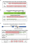
Fig. 1.
Fig. 1.
The generation of a Drosophila model of YPEL3 frameshift mutation. (A) CG15309 is the Drosophila homolog of human YPEL3. Sequence alignment between human YPEL3 (YPEL3) and Drosophila CG15309. Shaded in pink are the identical amino acid sequences. (B) Duplication of a cytosine nucleotide in YPEL3 gene from a patient (top). A predicted molecular lesion in human YPEL3 (bottom) introduces an ectopic amino acid sequence (shaded in green). The preserved region is shaded in pink. (C) CRISPR-Cas9 mediated in-del mutation in CG15309/dYPEL3. A guide RNA is designed targeting the middle of the coding exon (top). The isolated dYPEL3 in-del mutants (middle). Sequence alignment between wild type (wt), dYPEL3T1-6 and dYPEL3T1-8. The introduced ectopic amino acid sequences following a premature stop codon are shaded in green. The sequence alignment of the introduced ectopic amino acid sequences from the human YPEL3 frameshift mutants and dYPEL3T1-6 (bottom). The identical amino acid sequences are shaded in pink. ORF, open reading frame.
Fig. 2.
Fig. 2.
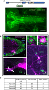
dYPEL3 is a neuronal gene. (A) The expression pattern of dYPEL3 in the CNS. The InSITE gene trap line for dYPEL3 was used (CG15309-GAL4/dYPEL3-GAL4). GAL4 transcription factor is inserted in the first intron. The introduction of UAS-mCD8::GFP demonstrates the endogenous expression pattern of dYPEL3. Note that the CG15309-GAL-positive cells elaborate fine processes throughout the CNS. Scale bar: 50 µm. (B) mCD8::GFP (magenta) was expressed under dYPEL3-GAL4 following immunostaining with anti-Elav (neuronal, green) and anti-Repo (glial, blue) antibodies. (i) The CNS. Cell bodies in 1 and 2 are shown magnified on the top right. (ii,iii) Chordotonal neurons (ii, arrows) and a class III da neuron (iii) in the PNS. iii also shows a class IV da neuron (nociceptor) that is positive for dYPEL3 (arrow). Scale bar: 10 µm. (C) Quantitation of the Elav-positive and Repo-positive cells that are labeled with CG15309-GAL4. The majority of dYPEL3-postive cells were Elav positive, but none were positive for Repo.
Fig. 3.
Fig. 3.
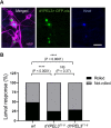
dYPEL3 frameshift mutations reduce nociceptive behavior. (A) Nociceptive/class IV da neurons are positive for dYPEL3. A nuclear GFP (GFP-nls, green) was expressed under dYPEL3-GAL4 following immunostaining with anti-Knot antibody (blue). Anti-HRP antibody was used to label all PNS neurons (magenta). Scale bar: 10 μm. (B) The AITC-induced nociceptive behavior was measured in a wild-type control (wt) and dYPEL3 frameshift mutants (dYPEL3T1-8 and dYPEL3T1-6). The number of larvae that exhibited complete rolling behavior was scored and expressed as a percentage (n=252 for each genotype). The Chi-squared test was performed between the groups. NS, non-significant; ****P<0.0001.
Fig. 4.
Fig. 4.
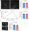
The development of nociceptors is not altered by dYPEL3 frameshift mutations. (A) mCD8::GFP was specifically expressed in nociceptors using ppk-GAL4 in wild-type control (wt) and dYPEL3 frameshift mutants (dYPEL3T1-8). Total length of dendrites was measured (n=6 for each genotype). Unpaired Student’s t-test with Welch's correction was performed. Scale bar: 50 µm. (B) Sholl analysis was performed with 20-µm radius increment from the dendritic tracing. Number of total crossings within 300 µm from the cell center was measured. Two-way ANOVA for Sholl analysis and unpaired Student's t-test with Welch's correction for total dendritic crossings were performed. (C) The axon terminals of single nociceptors from wild type and dYPEL3T1-8 mutants were visualized using the flip-out technique. The total length of axon terminals was measured (n=12 for wt, n=14 for dYPEL3T1-8). Scale bar: 10 µm. Unpaired Student's t-test with Welch's correction was performed. Data are presented as mean±s.e.m. All statistical analysis was two-tailed. NS, non-significant.
Fig. 5.
Fig. 5.
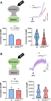
dYPEL3 frameshift mutations reduce the synaptic transmission from nociceptors to Basin-4 neurons. (A) Basin-4 activation upon AITC treatment was reduced by dYPEL3T1-8. GCaMP6f was expressed in Basin-4 neurons. Nociceptors were activated with 10 mM AITC (top left). Ca2+ increase in Basin-4 was measured by GCaMP fluorescence and the tracing over time is shown (n=40 for wt, n=47 for dYPEL3T1-8) (top right). The cumulative GCaMP activation from single Basin-4 neurons was measured and presented as mean±s.e.m. (bottom left), as well as in a violin plot to show distribution (bottom right). Mann–Whitney test. (B) Nociceptor activation was not altered by dYPEL3 mutations. GCaMP6f was expressed in nociceptors using ppk-GAL4. Nociceptors were activated with 10 mM AITC (top left). Ca2+ increase in the axon terminals of nociceptors was measured by GCaMP fluorescence and the tracing over time is shown (n=13 for each genotype) (top right). The cumulative GCaMP activation from the nociceptor axon terminals was measured and presented as mean±s.e.m. (bottom left), as well as in a violin plot to show distribution (bottom right). Mann–Whitney test. All statistical analysis was two-tailed. NS, non-significant; ****P<0.0001.
Fig. 6.
Fig. 6.
dYPEL3 frameshift mutations reduce the synaptic contact between nociceptors and Basin-4 neurons. (A) The syb-GRASP technique was used to report the synaptic contact between nociceptors and Basin-4. The spGFP1-10 (red cylinders) and spGFP11 (blue sectors) were expressed in nociceptors and Basin-4, respectively (left). The resulting GRASP signal was visualized by anti-GRASP antibody (green), and the spGFP1-10 that is expressed in nociceptor axon terminals was used as a normalization control (magenta) (right). Scale bar: 10 μm. (B) The GRASP intensity from each neuropil was normalized by spGFP1-10 intensity and presented as mean±s.e.m. (left), as well as in a violin plot to show distribution (right) (n=36 for wt, n=34 for dYPEL3T1-8). Mann–Whitney test. All statistical analysis was two-tailed. **P<0.01.
Fig. 7.
Fig. 7.
dYPEL3 knockout mutants recapitulate the phenotypes in the dYPEL3 frameshift mutants. (A) The generation of dYPEL3 knockout (dYPEL3KO) flies. Top: the entire dYPEL3 ORF was deleted using CRISPR/Cas9-mediated homology-directed recombination. Bottom: agarose gel image of PCR-based genotyping. Note that dYPEL3KO showed the PCR amplifications specific for DsRed-cassette, but not the ones from wild type. (B) dYPEL3 knockout reduces AITC-induced behavioral responses. AITC-induced nociceptive behavior was measured in a wild-type control (wt) and dYPEL3 knockout mutants (dYPEL3KO). The number of larvae that exhibited complete rolling behavior was scored and expressed as a percentage (n=252 for wt, n=203 for dYPEL3KO). The Chi-squared test was performed between the groups. (C) dYPEL3 knockout caused a reduction in Basin-4 activation upon AITC. GCaMP6f was expressed in Basin-4 neurons. Nociceptors were activated with 10 mM AITC. Ca2+ increase in Basin-4 was measured by GCaMP fluorescence and the GCaMP trace over time is shown (n=47 for wt, n=46 for dYPEL3KO) (left). The cumulative GCaMP activation from single Basin-4 neurons was measured and presented as mean±s.e.m. (middle), as well as in a violin plot to show distribution (right). Mann–Whitney test. (D) dYPEL3 knockout causes a reduction in syb-GRASP signals between nociceptors and Basin-4. Synaptobrevin-spGFP1-10 and spGFP11 were expressed in nociceptors and Basin-4, respectively. The resulting GRASP signal was visualized by anti-GFP antibody that only recognizes the reconstituted GFP (anti-GRASP) (green), and the spGFP1-10 expressed in nociceptor axon terminals was used as a normalization control (magenta) (left). Scale bar: 10 μm. The GRASP intensity from each neuropil was normalized by spGFP1-10 intensity and presented as mean±s.e.m. (middle), as well as in a violin plot to show distribution (right) (n=32 for wt, n=80 for dYPEL3T1-8). Mann–Whitney test. ***P<0.001 and ****P<0.0001.
Fig. 8.
Fig. 8.
The nociceptor-specific expression of dYPEL3 in dYPEL3 frameshift mutant rescues AITC-mediated larva rolling behavior. AITC-induced nociceptive behavior was measured. The nociceptor-specific expression of dYPEL3 was achieved using ppk-GAL. The number of larvae that exhibited complete rolling behavior was scored and expressed as a percentage (n=118 for wt, ppk>; n=115 for wt, ppk>dYPEL3; n=163 for dYPEL3T1-8, ppk>; n=103 for dYPEL3T1-8, ppk>dYPEL3). The Chi-squared test was performed between the groups. NS, non-significant; *P<0.05.

References

    1. Baker S. J. (2003). Small unstable apoptotic protein, an apoptosis-associated protein, suppresses proliferation of myeloid cells. Cancer Res. 63, 705-712. - PubMed
    1. Bellen H. J., Wangler M. F. and Yamamoto S. (2019). The fruit fly at the interface of diagnosis and pathogenic mechanisms of rare and common human diseases. Hum. Mol. Genet. 28, R207-R214. 10.1093/hmg/ddz135 - DOI - PMC - PubMed
    1. Bischof J., Maeda R. K., Hediger M., Karch F. and Basler K. (2007). An optimized transgenesis system for Drosophila using germ-line-specific C31 integrases. Proc. Natl. Acad. Sci. USA 104, 3312-3317. 10.1073/pnas.0611511104 - DOI - PMC - PubMed
    1. Blaker-Lee A., Gupta S., McCammon J. M., De Rienzo G. and Sive H. (2012). Zebrafish homologs of genes within 16p11.2, a genomic region associated with brain disorders, are active during brain development, and include two deletion dosage sensor genes. Dis. Model. Mech. 5, 834-851. 10.1242/dmm.009944 - DOI - PMC - PubMed
    1. Chang Y.-F., Imam J. S. and Wilkinson M. F. (2007). The nonsense-mediated decay RNA surveillance pathway. Annu. Rev. Biochem. 76, 51-74. 10.1146/annurev.biochem.76.050106.093909 - DOI - PubMed

Publication types

MeSH terms