. 2020 May;8(1):e000649.
doi: 10.1136/jitc-2020-000649.

Tumor ablation plus co-administration of CpG and saponin adjuvants affects IL-1 production and multifunctional T cell numbers in tumor draining lymph nodes

Affiliations

Tumor ablation plus co-administration of CpG and saponin adjuvants affects IL-1 production and multifunctional T cell numbers in tumor draining lymph nodes

Tonke K Raaijmakers et al. J Immunother Cancer. 2020 May.

Abstract

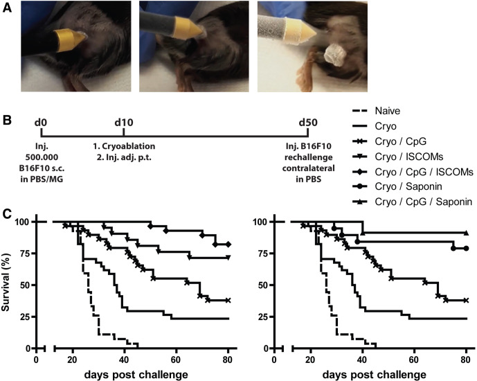
Background: Tumor ablation techniques, like cryoablation, are successfully used in the clinic to treat tumors. The tumor debris remaining in situ after ablation is a major antigen depot, including neoantigens, which are presented by dendritic cells (DCs) in the draining lymph nodes to induce tumor-specific CD8+ T cells. We have previously shown that co-administration of adjuvants is essential to evoke strong in vivo antitumor immunity and the induction of long-term memory. However, which adjuvants most effectively combine with in situ tumor ablation remains unclear.

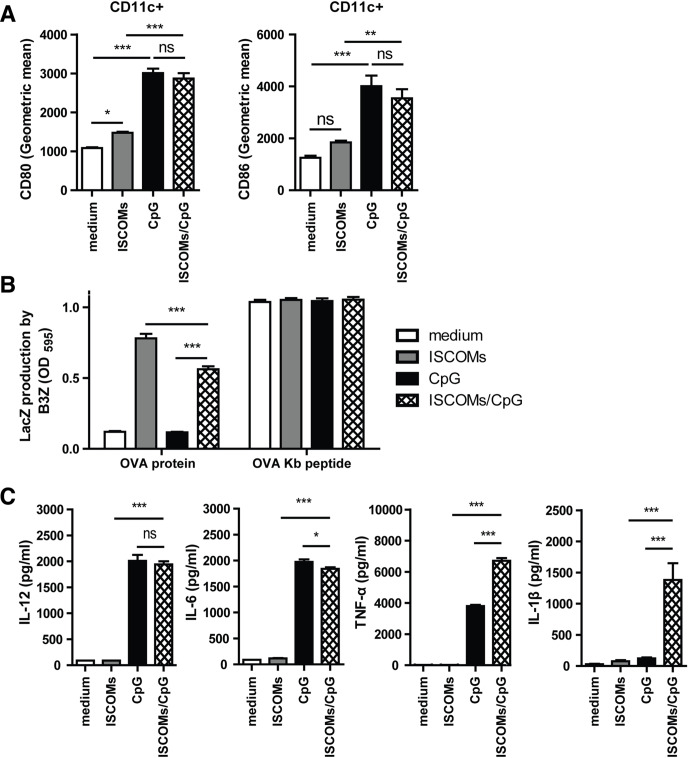
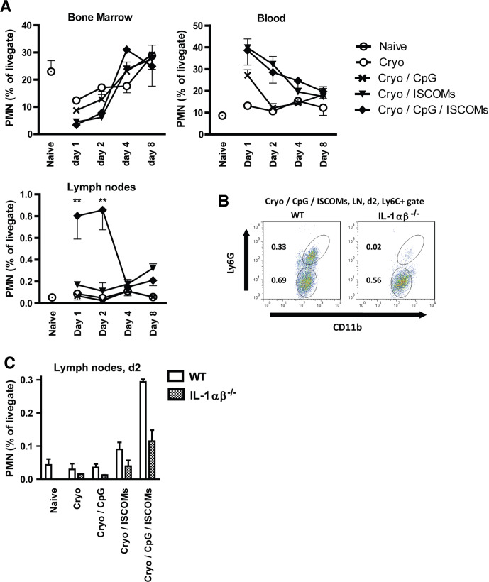
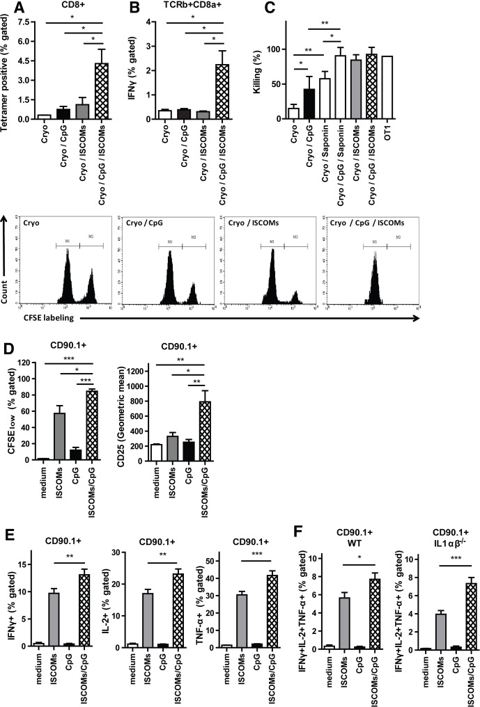
Methods and results: Here, we show that simultaneous administration of cytidyl guanosyl (CpG) with saponin-based adjuvants following cryoablation affects multifunctional T-cell numbers and interleukin (IL)-1 induced polymorphonuclear neutrophil recruitment in the tumor draining lymph nodes, relative to either adjuvant alone. The combination of CpG and saponin-based adjuvants induces potent DC maturation (mainly CpG-mediated), antigen cross-presentation (mainly saponin-based adjuvant mediated), while excretion of IL-1β by DCs in vitro depends on the presence of both adjuvants. Most strikingly, CpG/saponin-based adjuvant exposed DCs potentiate antigen-specific T-cell proliferation resulting in multipotent T cells with increased capacity to produce interferon (IFN)γ, IL-2 and tumor necrosis factor-α in vitro. Also in vivo the CpG/saponin-based adjuvant combination plus cryoablation increased the numbers of tumor-specific CD8+ T cells showing enhanced IFNγ production as compared with single adjuvant treatments.

Conclusions: Collectively, these data indicate that co-injection of CpG with saponin-based adjuvants after cryoablation induces an increased amount of tumor-specific multifunctional T cells. The combination of saponin-based adjuvants with toll-like receptor 9 adjuvant CpG in a cryoablative setting therefore represents a promising in situ vaccination strategy.

Keywords: CD8-positive T-lymphocytes; adaptive immunity; adjuvants, immunologic; dendritic cells; immunomodulation.


Conflict of interest statement

Competing interests: None declared.

Figures

Figure 1
Figure 1
Potent antitumor memory response following ablation combined with saponin-based adjuvants and CpG-ODN. (A) Visual representation of cryoablation on 6–8 mm tumors. (B) Established B16F10 tumors subcutaneously on the right femur were treated with cryoablation alone, or in combination with peritumoral injection of the indicated adjuvants (CpG-ODN and ISCOMs 30 µg, Quil A saponin 20 µg), within 30 min after ablation. Forty days later, naïve and tumor-free mice received a re-challenge with tumor cells subcutaneously on the flank. (C) Growth of this re-challenge is depicted as a Kaplan-Meier survival curve demonstrating superior protection from tumor outgrowth after combination of ablation with saponin-based adjuvants Quil A saponin or ISCOMs and CpG-ODN. Data pooled from three to four independent experiments, with 19–34 mice per group in total. Statistical significance was calculated using a log-rank test with post hoc Bonferroni correction. *P<0.05 for Cryo/CpG vs Cryo/CpG/ISCOMs and Cryo/CpG/saponin at day 80. Cryo, cryoablation; CpG, cytidyl guanosyl; ISCOMs, immune-stimulating complexes.
Figure 2
Figure 2
Combination of CpG and ISCOMs does not increase maturation, nor cross-presentation of antigens, but does affect cytokine production in vitro. Bone marrow DC were cultured with GM-CSF, and 2×10⁵ (A), 0.8×10⁵ (B) or 1–2×10⁵ (C) cells were exposed to 400 ng/mL ISCOMs and/or 1 µg/mL CpG-ODN and (B) 80 µg/mL endotoxin-free ovalbumin (OVA) or OVA Kb peptide. (A) After 16–24 hours, CD80 and CD86 expression was determined in CD11c+ population by flow cytometry (n=5). (B) After 5 hours stimulation, cells were washed and cultured overnight with 0.8×10⁵ B3Z cells, which produce LacZ on TCR recognition of OVA peptide in the context of H-2Kb. Next, LacZ production by the B3Z cells, as a measure of cross-presentation, was evaluated using a colorimetric assay (n=3). (C) After 16–24 hours, supernatant from GM-CSF DCs was harvested and the cytokines IL-6 (n=3), IL-12 (n=3), TNF-α (n=3) and IL-1β (n=21) were measured by ELISA. Results are shown as means with SEM. Statistical significance was calculated using a one-way analysis of variance with Tukey multiple comparison correction. *P<0.05; **p<0.01; ***p<0.001. CpG, cytidyl guanosyl; DC, dendritic cell; GM-CSF, granulocyte-macrophage colony-stimulating factor; IL, interleukin; ISCOMs, immune-stimulating complexes; ns, not significant; TNF, tumor necrosis factor.
Figure 3
Figure 3
CpG/ISCOMs causes IL-1 release, as reflected by PMN influx. (A–C) Established B16F10 tumors subcutaneously on the right femur of WT (A–C) and IL-1αβ-/- (B, C) mice were left untreated or cryoablated alone, or in combination with peritumoral injection of indicated adjuvants, within 30 min after ablation. After indicated time points (A, n>4) and 2 days postablation (B, C, n=2–3), bone marrow, blood, spleens (B) and draining lymph nodes (A–C) were collected and percentage of PMNs (Ly6C+Ly6G+CD11b+) were detected by flow cytometry. Results are shown as means with SEM. Statistical significance was calculated using a one-way analysis of variance with Tukey multiple comparison correction. **P<0.01. CpG, cytidyl guanosyl; IL, interleukin; ISCOMs, immune-stimulating complexes; PMN, polymorphonuclear neutrophil; WT, wild-type.
Figure 4
Figure 4
CpG/ISCOMs adjuvant treatment to DCs results in multifunctional T cells. (A–C) Established B16OVA tumors were treated with cryoablation alone, or in combination with the indicated adjuvants. (A) Seven days after ablation, lymph node cells were stained for OVA-Kb tetramers. (B) Seven days after ablation, cells from draining lymph nodes were restimulated with OVA Kb peptide and brefeldin A for 5 hours and TCR-β+CD8α+ cells were analyzed for the expression of IFNγ. (A, B) Results are shown as means with SEM (n=2). (C) After 10 days, the cryo-ablated and adjuvant injected mice, or one OT-I mouse, received CFSEhigh splenocytes pulsed with the target peptide (OVA Kb peptide), along with CFSElow splenocytes pulsed with the control peptide (HPV Kb peptide), in a ratio of 1:1. Inguinal lymph nodes of these mice were harvested 20 hours later and the relative numbers of CFSEhigh and CFSElow target cells were determined by flow cytometry. Data depicted are mean percentages of target cell killing (n=6 mice) with SD, corrected for background cell-death in naïve mice, and representative plots. Bone marrow DCs were cultured with GM-CSF, and 0.1×10⁵ (D) or 0.5×10⁵ (E, F) cells were exposed to 400 ng/mL ISCOMs and/or 1 µg/mL CpG-ODN and 80 µg/mL endotoxin-free ovalbumin (OVA protein). After 5 hours, cells were washed and cultured with 50×103 CFSE-labeled CD8+ T cells for 72 hours (D) or 1.2×10⁵ CD8+ OT-I T cells with brefeldin A overnight (E, F). (D) Cells were stained for CD90.1 and CD25. The expression of CD25 and the CFSE labeling in CD90.1+ population as a measure of proliferation were assessed by flow cytometry (n=3). (E, F) Intracellular build-up of IFNγ, IL-2 and TNF-α was measured using flow cytometry (E, WT, n=9 and F, IL-1ɑβ-/-, n=4). (D–F) Results are shown as means with SEM. (A–F) Statistical significance was calculated using a one-way analysis of variance with Tukey multiple comparison correction. *P<0.05; **p<0.01; ***p<0.001. CpG, cytidyl guanosyl; DC, dendritic cell; GM-CSF, granulocyte-macrophage colony-stimulating factor; IFN, interferon; IL, interleukin; ISCOMs, immune-stimulating complexes; TNF, tumor necrosis factor; WT, wild-type.

Similar articles

Cited by

References

    1. Sabel MS. Cryo-immunology: a review of the literature and proposed mechanisms for stimulatory versus suppressive immune responses. Cryobiology 2009;58:1–11. 10.1016/j.cryobiol.2008.10.126 - DOI - PubMed
    1. Yrlid U, Wick MJ. Antigen presentation capacity and cytokine production by murine splenic dendritic cell subsets upon Salmonella encounter. J Immunol 2002;169:108–16. 10.4049/jimmunol.169.1.108 - DOI - PubMed
    1. den Brok MHMGM, Sutmuller RPM, van der Voort R, et al. . In situ tumor ablation creates an antigen source for the generation of antitumor immunity. Cancer Res 2004;64:4024–9. 10.1158/0008-5472.CAN-03-3949 - DOI - PubMed
    1. den Brok MHMGM, Sutmuller RPM, Nierkens S, et al. . Efficient loading of dendritic cells following cryo and radiofrequency ablation in combination with immune modulation induces anti-tumour immunity. Br J Cancer 2006;95:896–905. 10.1038/sj.bjc.6603341 - DOI - PMC - PubMed
    1. Goldszmid RS, Idoyaga J, Bravo AI, et al. . Dendritic cells charged with apoptotic tumor cells induce long-lived protective CD4+ and CD8+ T cell immunity against B16 melanoma. J Immunol 2003;171:5940–7. 10.4049/jimmunol.171.11.5940 - DOI - PubMed

Publication types

MeSH terms