. 2020 Jun 9;117(23):12657-12664.
doi: 10.1073/pnas.2000605117. Epub 2020 May 27.

Semisynthesis of an evasin from tick saliva reveals a critical role of tyrosine sulfation for chemokine binding and inhibition

Affiliations

Semisynthesis of an evasin from tick saliva reveals a critical role of tyrosine sulfation for chemokine binding and inhibition

Charlotte Franck et al. Proc Natl Acad Sci U S A. .

Abstract

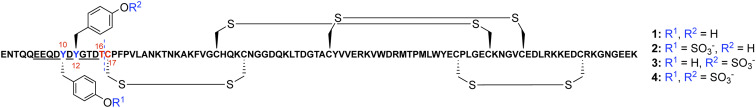
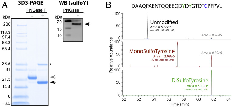
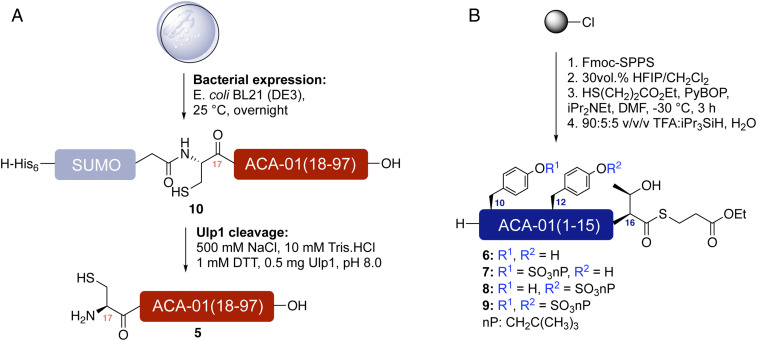
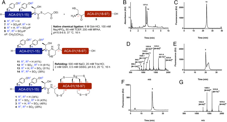
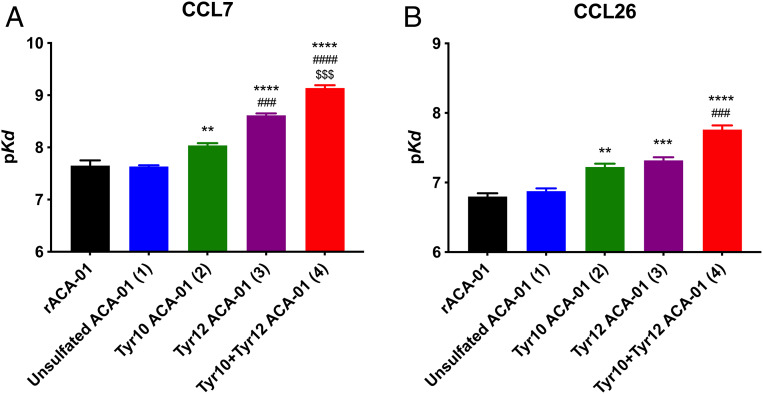
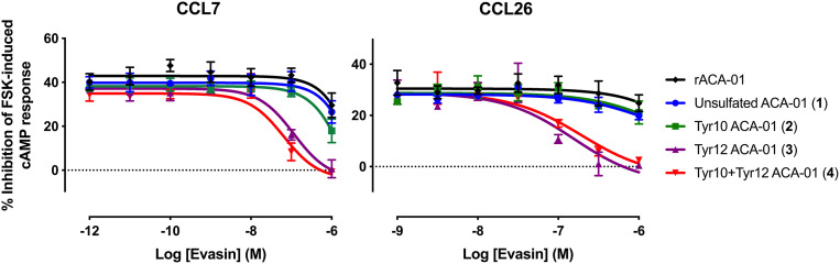
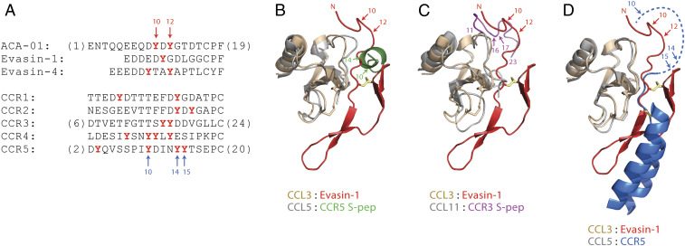
Blood-feeding arthropods produce antiinflammatory salivary proteins called evasins that function through inhibition of chemokine-receptor signaling in the host. Herein, we show that the evasin ACA-01 from the Amblyomma cajennense tick can be posttranslationally sulfated at two tyrosine residues, albeit as a mixture of sulfated variants. Homogenously sulfated variants of the proteins were efficiently assembled via a semisynthetic native chemical ligation strategy. Sulfation significantly improved the binding affinity of ACA-01 for a range of proinflammatory chemokines and enhanced the ability of ACA-01 to inhibit chemokine signaling through cognate receptors. Comparisons of evasin sequences and structural data suggest that tyrosine sulfation serves as a receptor mimetic strategy for recognizing and suppressing the proinflammatory activity of a wide variety of mammalian chemokines. As such, the incorporation of this posttranslational modification (PTM) or mimics thereof into evasins may provide a strategy to optimize tick salivary proteins for antiinflammatory applications.

Keywords: antiinflammatory; chemokines; evasins; sulfation; ticks.


Conflict of interest statement

The authors declare no competing interest.

Figures

Fig. 1.
Fig. 1.
Schematic of the ACA-01 evasin sequence. Two predicted Tyr sulfation sites (Y10 and Y12) are shown in blue flanked by a highly acidic stretch of amino acids from E6–D15 (underlined). T16–C17 (shown in red) was selected as the site for semisynthetic assembly of the ACA-01 sulfoproteins by native chemical ligation. The disulfide bond connectivity is predicted based on conservation of cysteine positioning with evasin-1 where disulfide bond connectivity has been confirmed through X-ray crystallography (25).
Fig. 2.
Fig. 2.
Tyrosine sulfation of evasin ACA-01 secreted by eukaryotic cells. (A) SDS-PAGE and Western blot analysis of purified ACA-01 secreted by HEK293 cells, before (−) and after (+) treatment with PNGase F to remove N-glycans. Gray and black arrows indicate N-glycosylated ACA-01-Myc-His6 and deglycosylated ACA-01-Myc-His6, respectively. PNGase F is indicated by an asterisk. (Left) Total protein was detected by Coomassie staining. (Right) Proteins containing sulfotyrosine were detected with immunoblotting using a pan-specific antisulfotyrosine antibody (see SI Appendix, Fig. S29 for full Western blot including nonsulfated ACA-01 as a negative control). (B) LC-MS/MS analysis of chymotryptic peptides derived from a digest of ACA-01 expressed in HEK293 cells. Extracted ion chromatograms were generated for the peptide sequence shown above that was modified at the tyrosine residues (green in sequence) with either both sulfated (green), only one sulfated (brown), or neither being modified (black). The peak area is shown for each ion type. The disulfotyrosine ion showed some artifactual desulfation during the process of ESI and ion transfer and the area of these peaks is shown.
Fig. 3.
Fig. 3.
Expression and synthesis of ACA-01 fragments. (A) Strategy for the generation of ACA-01(17–97) (5) through the expression of a His6-SUMO-ACA-01(17–97) (10) fusion followed by proteolysis with Ulp1. (B) Synthesis of ACA-01(1–15) (sulfo)peptide thioesters fragments (69) via Fmoc-strategy SPPS.
Fig. 4.
Fig. 4.
Assembly of differentially sulfated ACA-01 via native chemical ligation. (A) Semisynthesis of ACA-01 evasin (sulfo)proteins 14 via native chemical ligation and oxidative folding. Exemplar data for doubly sulfated ACA-01 4. NB: nP = CH2C(CH3)3; (B) UPLC trace of crude reaction of 5 with doubly sulfated peptide thioester 9; (C) analytical HPLC chromatogram of purified ligation product 14; (D) ESI mass spectrum of purified ligation product 14; (E) analytical HPLC trace of crude folding reaction of 14 to afford 4; (F) analytical HPLC trace of purified folded doubly sulfated ACA-01 protein 4; (G) ESI mass spectrum of purified folded doubly sulfated ACA-01 protein 4.
Fig. 5.
Fig. 5.
Binding of semisynthetic ACA-01 (sulfo)proteins 14 to CC chemokines. Affinities (pKd) derived from the competitive fluorescence anisotropy data for binding of all ACA-01 isoforms (14) and recombinantly expressed ACA-01 (rACA-01). Each panel shows the binding affinity of all isoforms for one chemokine: CCL7 (A) and CCL26 (B). Data represent the average pKd ± SEM of values from three independent experiments, each recorded in duplicate. *, #, and $ indicate significant differences from unsulfated ACA-01, Tyr10 ACA-01, and Tyr12 ACA-01, respectively. Significance (one-way ANOVA) is shown as ** P < 0.01; ***, ###, $$$ P < 0.001; ****, ####, $$$$ P < 0.0001. See SI Appendix, Figs. S21 and S22 for binding data.
Fig. 6.
Fig. 6.
Inhibition of chemokine activity by ACA-01 proteins 14. Shown are the inhibition profiles of chemokine activity by ACA-01 (sulfo)proteins (14) and recombinantly expressed ACA-01 (rACA-01) at concentrations ranging from 1 pM to 1 µM or 1 nM to 1 µM for CCL26 (80 nM) or CCL7 (30 nM), respectively, acting at receptor CCR2 (CCL7) or CCR3 (CCL26) in FlpIn TREx HEK293 cells. Chemokine activity was detected as the capacity of the chemokine to inhibit forskolin-induced production of cAMP, as detected via the BRET sensor, CAMYEL; differentially sulfated ACA-01 proteins (14) inhibit the cAMP-inhibitory activity of chemokines. Data points represent the average ± SEM of three independent experiments, each conducted in duplicate.
Fig. 7.
Fig. 7.
Sequence and structural comparisons of tyrosine sulfation sites in evasins and chemokine receptors. (A) Aligned partial sequences of the N-terminal regions of selected evasins (Top) and human CC chemokine receptors (Bottom). Alignment is based on the structural overlay of chemokine ligands in structures of evasin-1 bound to CCL3 and CCR5 bound to 5P7-CCL5 (D). Potentially sulfated tyrosine residues are in red bold type. (BD) Partial structure of evasin-1 (residues 1–39 shown as red ribbons, with the first disulfide in yellow) bound to CCL3 (wheat ribbons, disulfides as sticks) (25) overlayed with (B) the structure of a CCR5-derived doubly sulfated peptide (CCR5 S-pep, green) bound to CCL5 (gray) (48), (C) the structure of a CCR3-derived doubly sulfated peptide (CCR3 S-pep, magenta) bound to CCL11 (gray) (47), and (D) the structure of CCR5 (transmembrane helices TM1 and TM7 shown as blue ribbons, with the connecting disulfide in yellow) bound to 5P7-CCL5 (gray) (25, 46). The dashed blue line indicates the N-terminal region of CCR5, which is not defined in the crystal structure. Numbered arrows indicate the known or likely positions of Tyr sulfation. The structural overlays are based on chemokine sequence alignments extending from the CC motif to end of the C-terminal α-helix. Disordered regions of chemokines and peptides are omitted for clarity.

Similar articles

Cited by

References

    1. Bonecchi R. et al. ., Chemokines and chemokine receptors: An overview. Front. Biosci. 14, 540–551 (2009). - PubMed
    1. Rollins B. J., Chemokines. Blood 90, 909–928 (1997). - PubMed
    1. Lloyd C., Chemokines in allergic lung inflammation. Immunology 105, 144–154 (2002). - PMC - PubMed
    1. Charo I. F., Peters W., Chemokine receptor 2 (CCR2) in atherosclerosis, infectious diseases, and regulation of T-cell polarization. Microcirculation 10, 259–264 (2003). - PubMed
    1. Epperson M. L., Lee C. A., Fremont D. H., Subversion of cytokine networks by virally encoded decoy receptors. Immunol. Rev. 250, 199–215 (2012). - PMC - PubMed

Publication types