

Bilateral serous choroidal detachment in brucellosis and its management and outcome: Literature review and case report
- PMID: 32461483
- PMCID: PMC7508090
- DOI: 10.4103/ijo.IJO_1418_19
Bilateral serous choroidal detachment in brucellosis and its management and outcome: Literature review and case report
Abstract
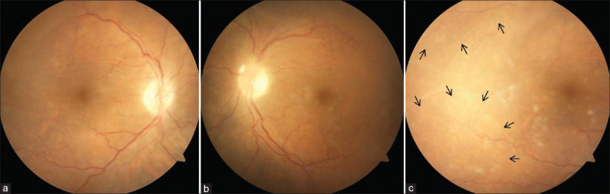
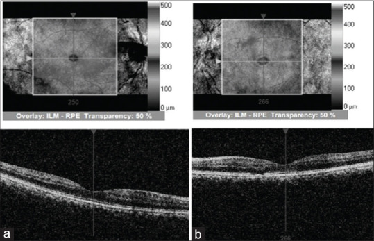
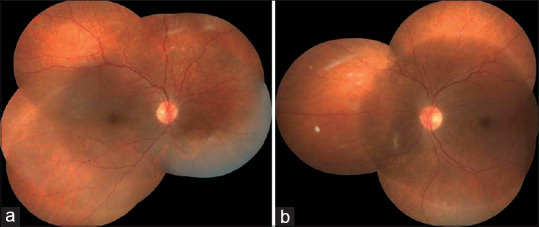
To report an unusual case of a 71-year-old livestock farmer with systemic brucellosis and ocular involvement. Examination showed vitreous haze with bilateral serous choroidal detachment. He was treated with topical antibiotics and corticosteroids, Tab rifampicin 600 mg and doxycycline 100 mg for 6 weeks with visual recovery and complete resolution of serous choroidal detachment in 2 weeks. This is the first case of bilateral serous choroidal detachment in a case of systemic brucellosis. Immune-mediated complex and direct microbial invasion of uveal tissue leading to serous choroidal detachment is the proposed pathogenesis that responds well to topical corticosteroids.
Keywords: Brucellosis; doxycycline; serous choroidal detachment.
Conflict of interest statement
None
Figures
Similar articles
-
PATIENT WITH UNILATERAL CHOROIDAL AND SEROUS RETINAL DETACHMENT WITH A HISTORY OF TREATED PROSTATE CANCER AND UNTREATED SARCOIDOSIS.Retin Cases Brief Rep. 2022 May 1;16(3):344-346. doi: 10.1097/ICB.0000000000000976. Epub 2020 Feb 13. Retin Cases Brief Rep. 2022. PMID: 32058354
-
Uveal Effusion Associated with Presumed Viral Encephalitis.Ocul Immunol Inflamm. 2022 Jan 2;30(1):68-72. doi: 10.1080/09273948.2020.1797110. Epub 2020 Aug 20. Ocul Immunol Inflamm. 2022. PMID: 32816570
-
Choroidal Effusion with Exudative Retinal Detachment following Non Perforating YAG-Laser Peripheral Iridotomy: A Case Report.Ocul Immunol Inflamm. 2024 Apr;32(3):358-361. doi: 10.1080/09273948.2023.2166850. Epub 2023 Jan 26. Ocul Immunol Inflamm. 2024. PMID: 36701763
-
Bilateral multifocal choroiditis with serous retinal detachment in a patient with Brucella infection: case report and review of the literature.Arch Ophthalmol. 2005 Jan;123(1):116-8. doi: 10.1001/archopht.123.1.116. Arch Ophthalmol. 2005. PMID: 15642826 Review. No abstract available.
-
Choroidal detachments: what do optometrists need to know?Clin Exp Optom. 2019 Mar;102(2):116-125. doi: 10.1111/cxo.12807. Epub 2018 Jul 4. Clin Exp Optom. 2019. PMID: 29971817 Review.
Cited by
-
Ocular Lesions in Brucella Infection: A Review of the Literature.Infect Drug Resist. 2022 Dec 22;15:7601-7617. doi: 10.2147/IDR.S394497. eCollection 2022. Infect Drug Resist. 2022. PMID: 36579126 Free PMC article. Review.
References
-
- Rolando I, Olarte L, Vilchez G, Lluncor M, Otero L, Paris M, et al. Ocular manifestations associated with brucellosis: A 26-year experience in Peru. Clin Infect Dis. 2008;46:1338–45. - PubMed
-
- Wechsler HF, Gutafson EG. Brucella endocarditis of congenital bicuspid aortic valve. Ann Intern Med. 1942;16:1228–33.
-
- Beebe RT, Meneely JK., Jr Brucella melitensis endocarditis; report of a case. Am Heart J. 8:788–91. 194. - PubMed
-
- Rabinowitz R, Schneck M, Levy J, Lifshitz T. Bilateral multifocal choroiditis with serous retinal detachment in a patient with Brucella infection: Case report and review of the literature. Arch Ophthalmol. 2005;123:116–8. - PubMed
-
- Woods A. Experimental Brucellar uveitis. Am J Ophthalmol. 1953;36:1025–42. - PubMed
Publication types
MeSH terms
LinkOut - more resources
Full Text Sources
Medical
