. 2020 May 27;11(1):2652.
doi: 10.1038/s41467-020-16488-y.

Prc1-rich kinetochores are required for error-free acentrosomal spindle bipolarization during meiosis I in mouse oocytes

Affiliations

Prc1-rich kinetochores are required for error-free acentrosomal spindle bipolarization during meiosis I in mouse oocytes

Shuhei Yoshida et al. Nat Commun. .

Abstract

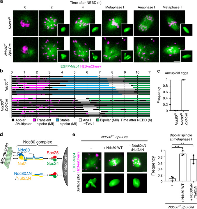
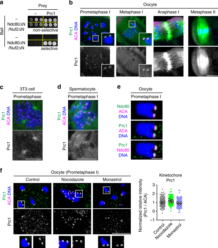
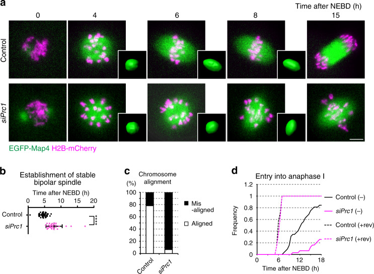
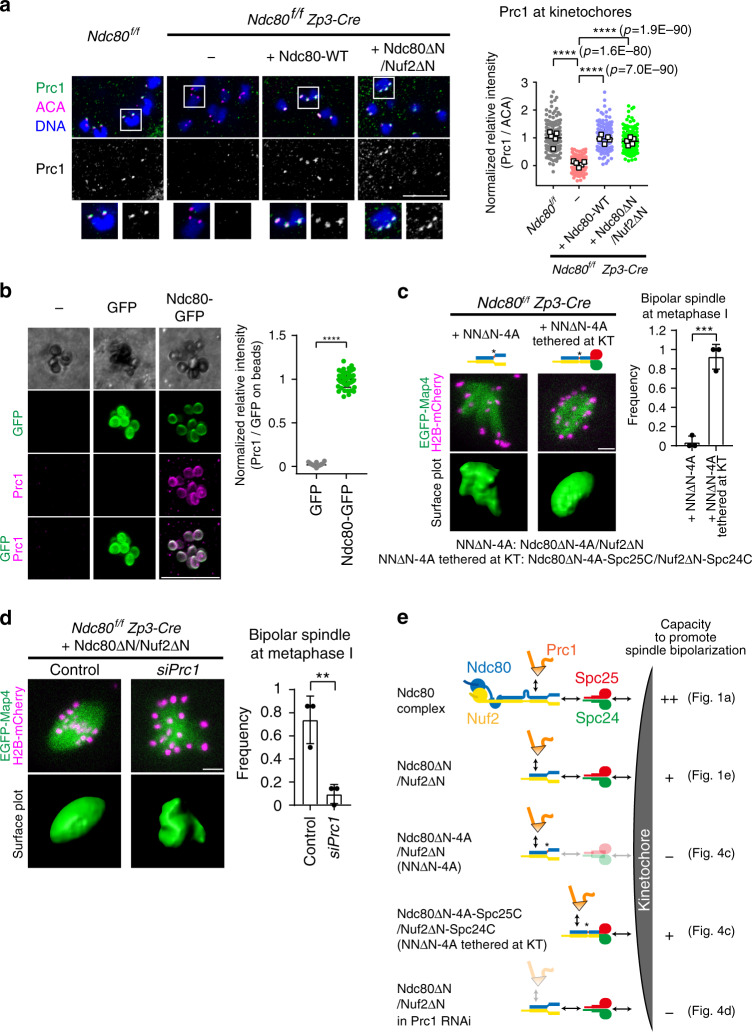
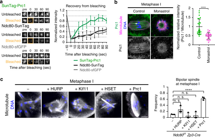
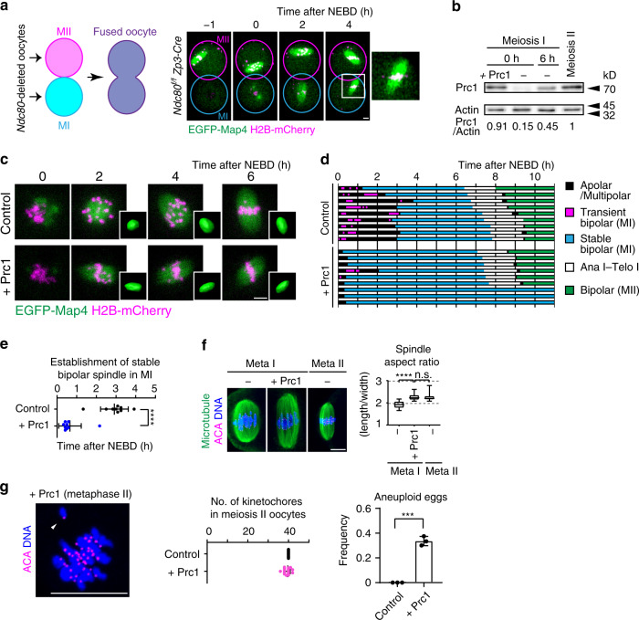
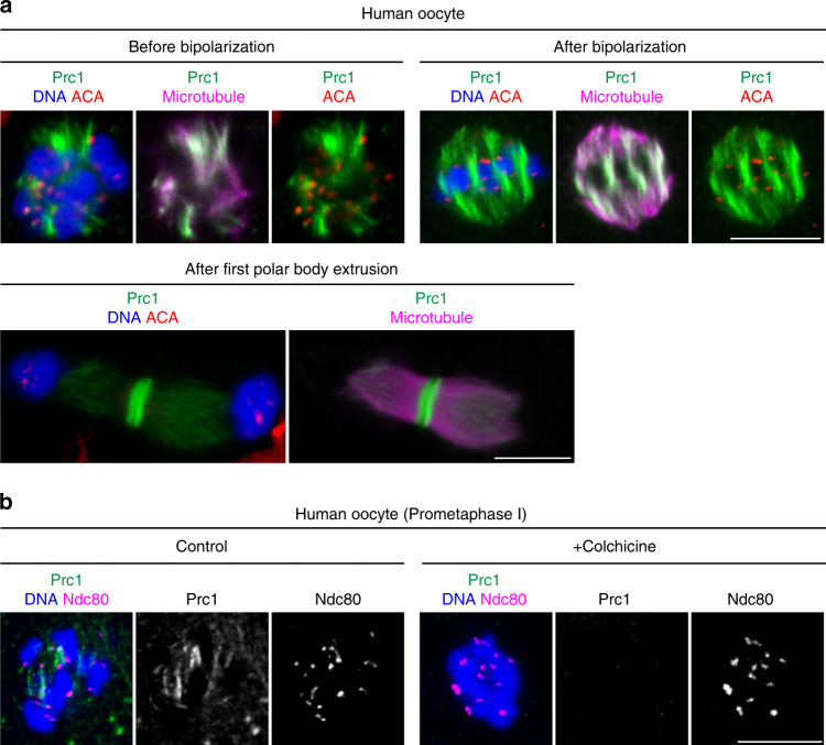
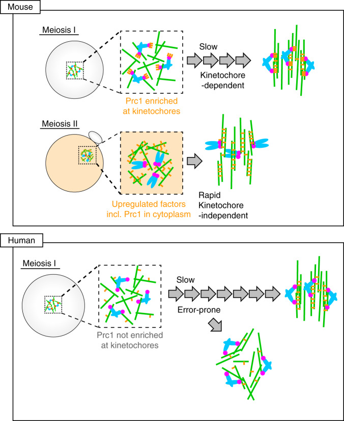
Acentrosomal meiosis in oocytes represents a gametogenic challenge, requiring spindle bipolarization without predefined bipolar cues. While much is known about the structures that promote acentrosomal microtubule nucleation, less is known about the structures that mediate spindle bipolarization in mammalian oocytes. Here, we show that in mouse oocytes, kinetochores are required for spindle bipolarization in meiosis I. This process is promoted by oocyte-specific, microtubule-independent enrichment of the antiparallel microtubule crosslinker Prc1 at kinetochores via the Ndc80 complex. In contrast, in meiosis II, cytoplasm that contains upregulated factors including Prc1 supports kinetochore-independent pathways for spindle bipolarization. The kinetochore-dependent mode of spindle bipolarization is required for meiosis I to prevent chromosome segregation errors. Human oocytes, where spindle bipolarization is reportedly error prone, exhibit no detectable kinetochore enrichment of Prc1. This study reveals an oocyte-specific function of kinetochores in acentrosomal spindle bipolarization in mice, and provides insights into the error-prone nature of human oocytes.


Conflict of interest statement

The authors declare no competing interests.

Figures

Fig. 1
Fig. 1. Ndc80 is essential for spindle bipolarization in MI but not in MII.
a Live imaging of Ndc80f/f (control) and Ndc80f/f Zp3-Cre (Ndc80-deleted) oocytes expressing EGFP-Map4 (microtubules, green) and H2B-mCherry (chromosomes, magenta). Spindle shapes were reconstructed in 3D. Asterisks show chromosomes that fell off the spindle. Anaphase I was defined based on characteristic chromosome and spindle dynamics (see Methods). Three independent experiments were performed. See also Supplementary Movie 1, 2, and 3. b Establishment of spindle bipolarity requires Ndc80 in MI. Spindle shapes in 3D were categorized based on the aspect ratio, surface irregularity, and stability (see Methods). Spindles that maintained a bipolar state for 1 h or longer with no collapse prior to anaphase I onset were categorized as stable bipolar spindles. c Ndc80 is required for preventing aneuploidy in eggs. The number of chromosomes were counted at metaphase II (n = 28, 28 oocytes from three independent experiments). Bars represent mean. See also Supplementary Movie 4. d The Ndc80 complex and Ndc80ΔN/Nuf2ΔN construct. In the Ndc80 complex, Ndc80 and Nuf2 directly bind to microtubules via the head domains at their N-termini, and to Spc24 and Spc25 at their C-termini. Ndc80 contains a loop domain in the central region, which recruits microtubule-binding proteins. Ndc80ΔN (a.a. 461–642) and Nuf2ΔN (a.a. 276–463) contain neither the head nor loop domain but retain the Spc24- and Spc25-binding domains. e Ndc80ΔN/Nuf2ΔN rescues spindle defects. Ndc80f/f Zp3-Cre oocytes coexpressing Ndc80ΔN and Nuf2ΔN (Ndc80ΔN/Nuf2ΔN) and those expressing full-length Ndc80 (Ndc80-WT) were monitored for spindle formation. EGFP-Map4 (microtubules, green) and H2B-mCherry (chromosomes, magenta) signals at metaphase I (5.5 h after NEBD) are shown. Spindle shapes were reconstructed in 3D at metaphase I (5.5 h after NEBD) and categorized based on the aspect ratio and surface irregularity (n = 23, 23, 21 oocytes from three independent experiments). Mean +/− SD are presented. **p = 0.0026; ***p = 0.0002; by two-tailed unpaired Student’s t-test. See also Supplementary Movie 5. Scale bars, 10 μm.
Fig. 2
Fig. 2. Ndc80/Nuf2 interacts with the antiparallel microtubule crosslinker Prc1 at kinetochores.
a Prc1 can interact with Ndc80ΔN/Nuf2ΔN. Yeast two-hybrid assay using Ndc80ΔN fused with a DNA-binding domain, Nuf2ΔN, and Prc1 fused with a transcription activation domain. Selective (−His, −Ade) and nonselective (+His, +Ade) plates were used. b Prc1 localizes to kinetochores. Z-projection images of oocytes stained for Prc1 (green), kinetochores (ACA, magenta), and DNA (Hoechst33342, blue) are shown. Prc1 signals at kinetochores are magnified. One of the z-slice images at metaphase II is shown in Supplementary Fig. 6g. More than three independent experiments were performed. c Prc1 is not enriched at kinetochores at mitotic prometaphase in centrosomal cultured cells. NIH3T3 cells were stained for Prc1 (green), kinetochores (ACA, magenta), and DNA (Hoechst33342, blue). Three independent experiments were performed. d Prc1 is not enriched at kinetochores at meiotic prometaphase I in centrosomal spermatocytes. Spermatocytes were stained for Prc1 (green), kinetochores (ACA, magenta), and DNA (Hoechst33342, blue). Three independent experiments were performed. e Prc1 colocalizes with Ndc80. Oocytes expressing Ndc80-sfGFP at prometaphase I (2 h after NEBD) were immunostained with anti-GFP, anti-Prc1, and ACA antibodies. DNA is counterstained with Hoechst33342 (blue). Magnified images show that Prc1 is located closer to Ndc80 than to ACA. Three independent experiments were performed. f Prc1 localizes to kinetochores independently of microtubule attachment and spindle bipolarization. Oocytes were treated with nocodazole or monastrol at 1.5 h after NEBD, incubated for 30 min, and immunostained for Prc1 (green), kinetochores (ACA, magenta), and DNA (Hoechst33342, blue). Prc1 signals at kinetochores are magnified. The kinetochore ratios of the Prc1 signals to the ACA signal are shown. Spots and squares correspond to individual kinetochores and oocytes, respectively (n = 200, 200, 198 kinetochores from 5, 5, 5 oocytes). Three independent experiments were performed. Mean +/− SD are presented. Scale bars; 10 μm (b), (c), (d), and (f); 2 μm (e).
Fig. 3
Fig. 3. Prc1 is required for timely spindle bipolarization in MI.
a Live imaging of oocytes after Prc1 RNAi. Ndc80f/f oocytes labeled with EGFP-Map4 (microtubules, green) and H2B-mCherry (chromosomes, magenta) were used. Spindle shapes were reconstructed in 3D. Four independent experiments were performed. Scale bar, 10 μm. See also Supplementary Movie 6. b Prc1 depletion delays spindle bipolarization. Spindle shapes in 3D were categorized based on the aspect ratio, surface irregularity, and stability (see Methods). The plot shows the time at which a stable bipolar spindle was established (n = 32, 29 oocytes from four independent experiments). Mean +/− SD are presented. ****p < 0.0001 (p = 2.4E−08) by two-tailed unpaired Student’s t-test. c Prc1 depletion increases chromosome misalignment. Oocytes were categorized into ‘aligned’ when all chromosomes located at the middle half of the spindle (n = 32, 31 oocytes from four independent experiments). d Prc1 depletion causes a delay in anaphase I onset. Oocytes were incubated with or without the checkpoint inhibitor reversine (rev). The percentages of oocytes that underwent anaphase I onset were plotted (n = 32, 31, 14, 14 oocytes from at least three independent experiments).
Fig. 4
Fig. 4. Ndc80/Nuf2 promotes spindle bipolarization by recruiting Prc1 at kinetochores.
a Ndc80/Nuf2 recruits Prc1 at kinetochores. Ndc80f/f (control) and Ndc80f/f Zp3-Cre (Ndc80-deleted) oocytes expressing full-length Ndc80 (Ndc80-WT) or Ndc80ΔN/Nuf2ΔN were immunostained for Prc1 (green), kinetochores (ACA, magenta), and DNA (Hoechst33342, blue). Kinetochores are magnified. In the plot, spots and squares correspond to individual kinetochores and oocytes, respectively (n = 200, 198, 200, 200 kinetochores from 5, 5, 5, 5 oocytes). ****p < 0.0001 (exact values are shown in the panel) by two-tailed unpaired Student’s t-test. b Ndc80-bound beads associate with Prc1. Oocytes expressing mEGFP or Ndc80-mEGFP (green) were microinjected with anti-GFP beads, fixed at prometaphase I, and immunostained for Prc1 (magenta) (see Methods). In the plot, spots correspond to individual beads (n = 50, 48 beads from 5, 5 oocytes). ****p < 0.0001 (p = 4.7E−87) by two-tailed unpaired Student’s t-test. c Ndc80/Nuf2–Prc1 acts at kinetochores. Ndc80f/f Zp3-Cre oocytes expressing Ndc80ΔN-4A/Nuf2ΔN (Y564A, Q565A, L566A, and T567A mutations; indicated as ‘NNΔN-4A’) or its Spc-fused form Ndc80ΔN-4A-Spc25C/Nuf2ΔN-Spc24C (indicated as ‘NNΔN-4A tethered at KTs’) were monitored. Spc25C (a.a. 120–226) and Spc24C (a.a. 122–201) are kinetochore-targeting domains. EGFP-Map4 (microtubules, green) and H2B-mCherry (chromosomes, magenta) at metaphase I (5.5 h after NEBD) are shown. Spindles were reconstructed in 3D and categorized based on the aspect ratio and surface irregularity. The frequency of oocytes that exhibited a bipolar-shaped spindle is shown (n = 19, 18 oocytes from three independent experiments). ***p = 0.0004 by two-tailed unpaired Student’s t-test. d Ndc80ΔN/Nuf2ΔN requires Prc1 to promote spindle bipolarization. Prc1 was depleted by RNAi in Ndc80f/f Zp3-Cre oocytes, and Ndc80ΔN/Nuf2ΔN was expressed. EGFP-Map4 (microtubules, green) and H2B-mCherry (chromosomes, magenta) at metaphase I (8 h after NEBD) are shown. Spindle shapes were reconstructed in 3D and categorized based on the aspect ratio and surface irregularity. The frequency of oocytes that exhibited a bipolar-shaped spindle is shown (n = 27, 27 oocytes from three independent experiments). **p = 0.0021 by two-tailed unpaired Student’s t-test. e Summary of Ndc80/Nuf2 constructs and their capacities to rescue spindle bipolarization. Scale bars, 10 μm. Mean +/− SD are presented in ad.
Fig. 5
Fig. 5. Ndc80/Nuf2 concentrates dynamic Prc1 at kinetochores for spindle bipolarization.
a Dynamic exchange of Prc1 at kinetochores. In oocytes at prometaphase I (1.5 h after NEBD), SunTag-Prc1 (24xGCN4-Prc1 coexpressed with scFv-sfGFP, green) signals at kinetochores were bleached, and the recovery was monitored (n = 5, 5, 5 kinetochores). Note that the recovery curve of SunTag-Prc1 signals indicates the turnover of Prc1 at kinetochores rather than the turnover of scFv on 24xGCN4 in this time range, which was confirmed by Ndc80-SunTag (Ndc80-24xGCN4 coexpressed with scFv-sfGFP) exhibiting similar recovery curves to Ndc80-sfGFP. See also Supplementary Movie 7. b Kif11-dependent Prc1 enrichment along kinetochore-proximal microtubules of the bipolar spindle. Control or monastrol-treated oocytes at metaphase I (4–6 h after NEBD) were stained for Prc1 (green), microtubules (magenta), and DNA (Hoechst33342, blue). The oocytes were treated with a cold buffer for 1 min before fixation to facilitate antibody penetration into the spindle. Prc1 signals along kinetochore-proximal microtubule bundles were measured, and their ratio to microtubule signals was calculated (n = 25, 25 locations from 5, 5 oocytes. Three independent experiments were performed). ****p < 0.0001 (p = 8.1E−07) by two-tailed unpaired Student’s t-test. c Prc1 overexpression rescues spindle defects in Ndc80-deleted oocytes. Ndc80f/f Zp3-Cre oocytes overexpressing mEGFP-HURP, mEGFP-Kif11, mNeonGreen-HSET, or mNeonGreen-Prc1 were immunostained at metaphase I (5.5 h after NEBD). Spindle shapes were reconstructed in 3D and categorized (n = 29, 18, 18, 18, 24 oocytes from three independent experiments). ****p < 0.0001 (p = 5.1E−05) by two-tailed unpaired Student’s t-test. n.s., not significant. Scale bars, 10 μm. Mean +/− SD are presented in ac.
Fig. 6
Fig. 6. Cytoplasm containing upregulated Prc1 facilitates kinetochore-independent pathways for spindle bipolarization in MII.
a MII cytoplasm supports Ndc80-independent spindle bipolarization. An Ndc80f/f Zp3-Cre oocyte in metaphase II (MII) and an Ndc80f/f Zp3-Cre oocyte at the GV stage (MI) were fused. EGFP-Map4 (microtubules, green) and H2B-mCherry (chromosomes, magenta) are shown. Bipolar spindle formation around MI chromosomes (magnified) was observed in 10 of 10 oocytes from three independent experiments. Time after NEBD of the MI nucleus (h). See also Supplementary Movie 8. b Upregulation of Prc1 in MII and artificial increase in Prc1 levels in MI. Western blotting of oocytes at MI (0 and 6 h after NEBD), and at MII (16 h). The Prc1 level at 0 h was artificially increased (+Prc1). Fifty oocytes were used in each sample. A full scan image is provided in the Source Data file. c Live imaging of oocytes expressing increased Prc1. EGFP-Map4 (microtubules, green) and H2B-mCherry (chromosomes, magenta) were monitored. Spindles were reconstructed in 3D. Time after NEBD. Three independent experiments were performed. See also Supplementary Movie 9. d Increased Prc1 accelerates spindle bipolarization. Spindles reconstructed in 3D were categorized (see Methods). e Acceleration of bipolar spindle establishment. Time at which a stable bipolar spindle was established are plotted (n = 9, 9 oocytes). The mean +/− SD are shown. ****p = 1.3E−06. f MII-like spindle shapes. Oocytes were immunostained for microtubules (green), kinetochores (ACA, magenta) and DNA (Hoechst33342, blue) at metaphase I (6 h after NEBD) and metaphase II (16 h after NEBD). Aspect ratios (length/width) were plotted (n = 19, 8, 11 oocytes). Boxes show the 25th to 75th percentiles and whiskers show the 1st to 99th percentiles. ****p = 2.54E−05. n.s., not significant. g Increased Prc1 increases aneuploidy in eggs. Oocytes at metaphase II were immunostained for kinetochores (ACA, magenta) and counterstained for DNA (Hoechst33342, blue). Arrowheads indicate a misaligned chromatid in an aneuploid egg. The number of kinetochores were counted (n = 33, 40 oocytes from three independent experiments). Mean +/− SD are presented. ***p = 0.0001. Statistical values were obtained from two-tailed unpaired Student’s test. Scale bars, 10 μm.
Fig. 7
Fig. 7. Human oocytes do not exhibit detectable enrichment of Prc1 at kinetochores.
a Prc1 localizes along spindle microtubules but not at kinetochores in human oocytes. MI oocytes of young women with no history of infertility were immunostained for Prc1 (green), kinetochores (ACA, red), microtubules (magenta) and DNA (DAPI, blue). Stages are categorized based on chromosome arrangement. Prc1 is detected at the spindle midzone after anaphase I. Oocytes from three women were examined. b Prc1 is not detectable at kinetochores. Human oocytes 5 h post NEBD were treated with the microtubule depolymerizer colchicine and immunostained for Prc1 (green), Ndc80 (red), and DNA (Hoechst33342, blue). Oocytes from more than three women were examined. Scale bars, 10 μm.
Fig. 8
Fig. 8. Models for acentrosomal spindle bipolarization in mammalian oocytes.
Different modes of spindle bipolarization. In mouse MI oocytes, Prc1 (orange) is highly enriched at kinetochores (magenta). The Prc1-rich kinetochore microenvironment is required for driving slow bipolarization of the spindle (green). This kinetochore-dependent spindle bipolarization is critical for MI to prevent chromosome segregation errors. In mouse MII oocytes, the cytoplasmic environment that contains upregulated factors including Prc1 provides global support to rapid, kinetochore-independent pathways for spindle bipolarization. In human MI oocytes, spindle bipolarization occurs without kinetochore-enriched Prc1, and is error prone.

Similar articles

Cited by

References

    1. Dumont J, Desai A. Acentrosomal spindle assembly and chromosome segregation during oocyte meiosis. Trends Cell Biol. 2012;22:241–249. - PMC - PubMed
    1. Bennabi I, Terret M-E, Verlhac M-H. Meiotic spindle assembly and chromosome segregation in oocytes. J. Cell Biol. 2016;215:611–619. - PMC - PubMed
    1. Mogessie, B., Scheffler, K. & Schuh, M. Assembly and positioning of the oocyte meiotic spindle. Annu. Rev. Cell Dev. Biol.34, 381–403 (2018). - PubMed
    1. Howe K, FitzHarris G. Recent insights into spindle function in mammalian oocytes and early embryos. Biol. Reprod. 2013;89:71. - PubMed
    1. Ohkura H. Meiosis: an overview of key differences from mitosis. Csh Perspect. Biol. 2015;7:a015859. - PMC - PubMed

Publication types