. 2020 Nov;33(11):2280-2294.
doi: 10.1038/s41379-020-0571-7. Epub 2020 May 27.

Invasive squamous cell carcinomas and precursor lesions on UV-exposed epithelia demonstrate concordant genomic complexity in driver genes

Affiliations

Invasive squamous cell carcinomas and precursor lesions on UV-exposed epithelia demonstrate concordant genomic complexity in driver genes

Lorena Lazo de la Vega et al. Mod Pathol. 2020 Nov.

Abstract

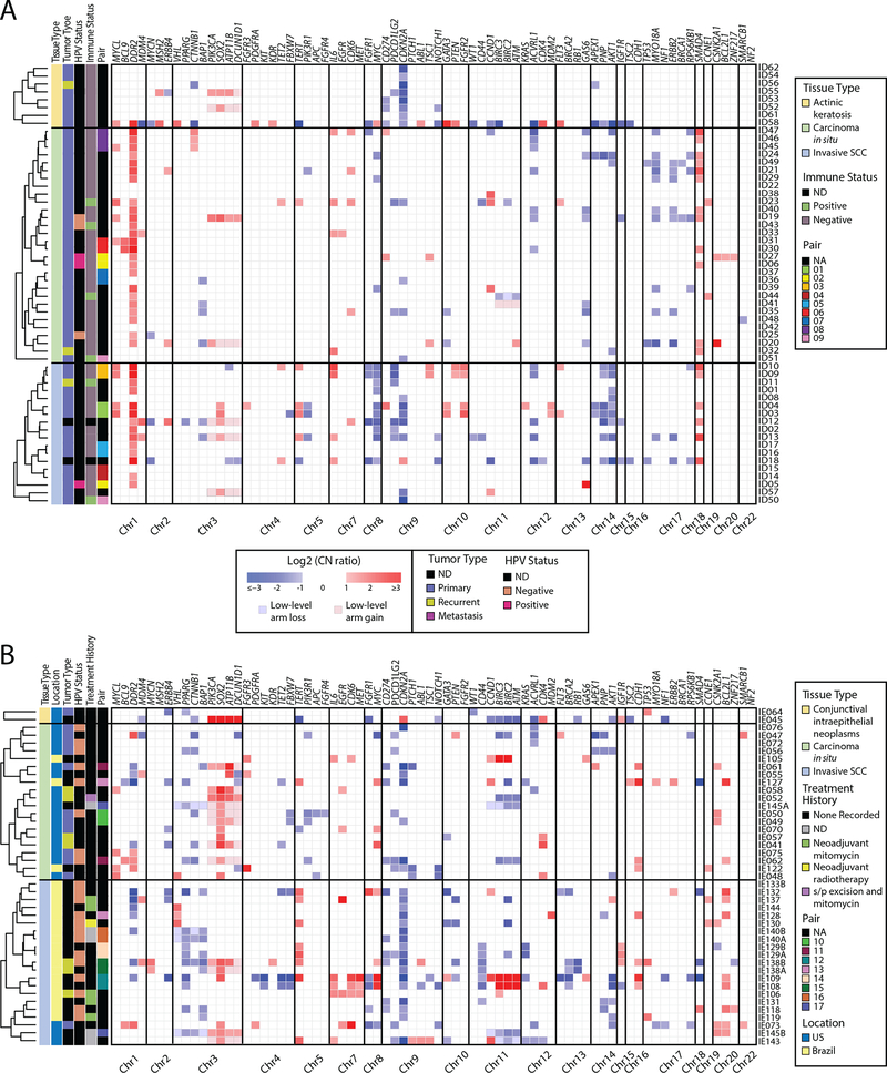
Although squamous cell carcinomas (SCC) are the most frequent human solid tumor at many anatomic sites, the driving molecular alterations underlying their progression from precursor lesions are poorly understood, especially in the context of photodamage. Therefore, we used high-depth, targeted next-generation sequencing (NGS) of RNA and DNA from routine tissue samples to characterize the progression of both well- (cutaneous) and poorly (ocular) studied SCCs. We assessed 56 formalin-fixed paraffin-embedded (FFPE) cutaneous lesions (n = 8 actinic keratosis, n = 30 carcinoma in situ [CIS], n = 18 invasive) and 43 FFPE ocular surface lesions (n = 2 conjunctival/corneal intraepithelial neoplasia, n = 20 CIS, n = 21 invasive), from institutions in the US and Brazil. An additional seven cases of advanced cutaneous SCC were profiled by hybrid capture-based NGS of >1500 genes. The cutaneous and ocular squamous neoplasms displayed a predominance of UV-signature mutations. Precursor lesions had highly similar somatic genomic landscapes to SCCs, including chromosomal gains of 3q involving SOX2, and highly recurrent mutations and/or loss of heterozygosity events affecting tumor suppressors TP53 and CDKN2A. Additionally, we identify a novel molecular subclass of CIS with RB1 mutations. Among TP53 wild-type tumors, human papillomavirus transcript was detected in one matched pair of cutaneous CIS and SCC. Amplicon-based whole-transcriptome sequencing of select 20 cutaneous lesions demonstrated significant upregulation of pro-invasion genes in cutaneous SCCs relative to precursors, including MMP1, MMP3, MMP9, LAMC2, LGALS1, and TNFRSF12A. Together, ocular and cutaneous squamous neoplasms demonstrate similar alterations, supporting a common model for neoplasia in UV-exposed epithelia. Treatment modalities useful for cutaneous SCC may also be effective in ocular SCC given the genetic similarity between these tumor types. Importantly, in both systems, precursor lesions possess the full complement of major genetic changes seen in SCC, supporting non-genetic drivers of invasiveness.


Conflict of interest statement

Conflict of Interest: S.A.T. has had a prior sponsored research agreement with ThermoFisher Scientific that provided access to the OCP. S.A.T. is a co-founder of, prior consultant to, equity holder in, and current employee of Strata Oncology. A.M.C. is a consultant and SAB member of Tempus.

Figures

FIGURE 1
FIGURE 1. Cutaneous squamous carcinoma and precursor lesions by light microscopy (left) and molecular features (right).
(A) Actinic keratosis displaying atypia of the basal layer of the epidermis, with maturation in the upper layers. Copy number profiling demonstrated CDKN2A loss. Nonsynonymous mutations including truncating mutation of TP53 and truncating mutations of CDKN2A. (B) Squamous cell in situ, demonstrating full-thickness squamous atypia without invasion. Molecular features include CDKN2A loss and mutation, accompanied by TP53 mutations. (C) Invasive squamous cell carcinoma adjacent to the in situ lesion in the panel above, displaying malignant squamous cells infiltrating collagen. Molecular findings include CDKN2A loss and distinct TP53 mutations.
FIGURE 2
FIGURE 2. Somatic copy number profiles of (A) cutaneous and (B) ocular lesions generated by targeted next generation sequencing (NGS).
Somatic, autosomal copy number profiles are presented for (A) 63 cutaneous and (B) 43 ocular tissues. Each copy number profile was GC and tumor content corrected. Normalized read counts per amplicon were divided by those from composite normal tissue, yielding a copy number ratio for each gene (cancer/composite normal), with red and blue indicating gain and loss, respectively, according to the log2 color scale (right). Unsupervised clustering was used on all log2 copy number ratios within lesion groups. Copy number ratios between the range of −1 and 1 were not visualized. Genes part of low arm-level gains and losses are shown with a different shade and border. Columns represent individual targeted genes in genome order (from chromosome 1 to 22). Clinicopathologic features are indicated in the figure legend. MI-Oncoseq cases are not shown due to differences in normalization. ND: Not Determined; NA: Not Available
FIGURE 3
FIGURE 3. Integrated heatmap of prioritized mutations and copy number aberrations identified by next generation sequencing.
Integrated table of prioritized nonsynonymous mutations and copy number aberrations from (A) 63 cutaneous and (B) 43 ocular tissues. Rows represent genes and columns represent individual samples. Clinicopathologic features are indicated in the figure legend. Copy number aberrations and prioritized mutation types are indicated below the table. A total of eight ocular tissues samples were only analyzed for copy number aberrations and not for mutations, as labeled. Thresholds used: Loss (1-copy loss), Deep deletion (2-copy loss), Gain (1 or 2 copy gain), Amplification (>2-copy gain)
FIGURE 4
FIGURE 4. Two-level concentric pie charts and CDKN2A variant mapping.
Two-level concentric pie charts show zygosity (each level) and co-occurrence (overlapping regions of the two levels) of CDKN2A mutations and CDKN2A variant mapping across (A) AK, CIS, invasive cutaneous SCC and B) CIS and invasive conjunctival SCC. Outside circle gives the number of samples with a homozygous (Homo) mutations. Inside circle gives the number of samples with heterozygous (Het) mutations. The number of heterozygous/homozygous CDKN2A mutations in each section is denoted by shading. Overlapping regions of the pie chart indicate samples with multiple mutations at homozygous and heterozygous mutations. CDKN2A (NM_000077) mutations in cutaneous and ocular SCCs were arranged by amino acid location. Histological classification is noted by the color of each post segment. Mutation type is labeled by the colored dot as according to the figure legend.
FIGURE 5
FIGURE 5. Gene expression heatmap generated from cutaneous SCC whole-transcriptome amplicon-based RNA-seq.
Heatmap of median-centered expression of 129 overlapping differentially expressed genes from the AK versus invasive SCC and in situ versus cutaneous invasive SCC comparison. Clinicopathologic features are indicated in the figure legend. ND: Not Determined; NA: Not Available

Similar articles

Cited by

References

    1. Dotto GP, Rustgi AK. Squamous Cell Cancers: A Unified Perspective on Biology and Genetics. Cancer Cell, 2016. 29: 622–37. - PMC - PubMed
    1. Campbell JD, Yau C, Bowlby R, Liu Y, Brennan K, Fan H, et al. Genomic, Pathway Network, and Immunologic Features Distinguishing Squamous Carcinomas. Cell Rep, 2018. 23: 194–212 e6. - PMC - PubMed
    1. Pickering CR, Zhou JH, Lee JJ, Drummond JA, Peng SA, Saade RE, et al. Mutational landscape of aggressive cutaneous squamous cell carcinoma. Clin Cancer Res, 2014. 20: 6582–92. - PMC - PubMed
    1. Li YY, Hanna GJ, Laga AC, Haddad RI, Lorch JH, Hammerman PS. Genomic analysis of metastatic cutaneous squamous cell carcinoma. Clin Cancer Res, 2015. 21: 1447–56. - PMC - PubMed
    1. Inman GJ, Wang J, Nagano A, Alexandrov LB, Purdie KJ, Taylor RG, et al. The genomic landscape of cutaneous SCC reveals drivers and a novel azathioprine associated mutational signature. Nat Commun, 2018. 9: 3667. - PMC - PubMed

Publication types

MeSH terms