. 2020 May 12:2020:3214186.
doi: 10.1155/2020/3214186. eCollection 2020.

α 1-Adrenergic Receptor Blockade by Prazosin Synergistically Stabilizes Rat Peritoneal Mast Cells

Affiliations

α 1-Adrenergic Receptor Blockade by Prazosin Synergistically Stabilizes Rat Peritoneal Mast Cells

Nozomu Abe et al. Biomed Res Int. .

Retraction in

Abstract

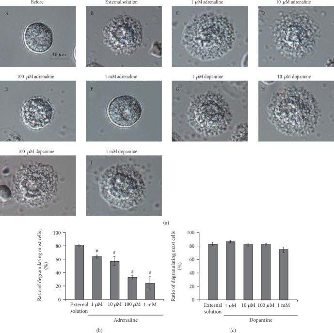
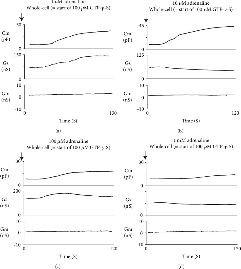
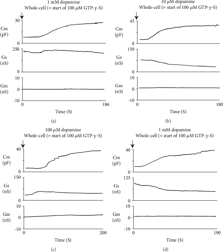
Background: Adrenaline quickly inhibits the release of histamine from mast cells. Besides β 2-adrenergic receptors, several in vitro studies also indicate the involvement of α-adrenergic receptors in the process of exocytosis. Since exocytosis in mast cells can be detected electrophysiologically by the changes in the membrane capacitance (Cm), its continuous monitoring in the presence of drugs would determine their mast cell-stabilizing properties.

Methods: Employing the whole-cell patch-clamp technique in rat peritoneal mast cells, we examined the effects of adrenaline on the degranulation of mast cells and the increase in the Cm during exocytosis. We also examined the degranulation of mast cells in the presence or absence of α-adrenergic receptor agonists or antagonists.

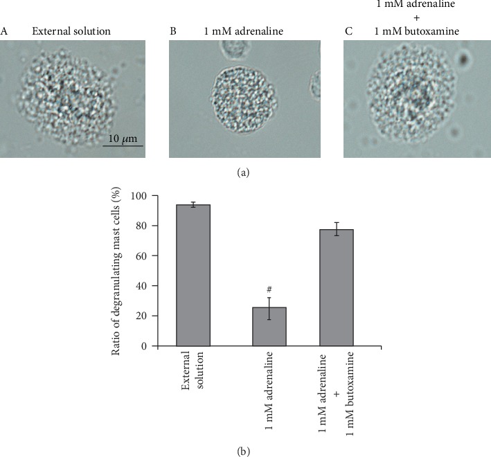
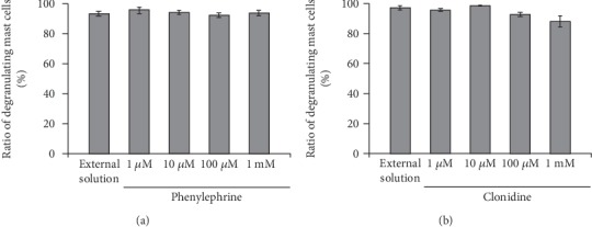
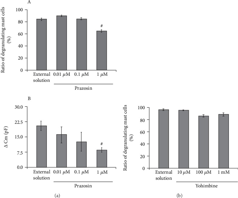
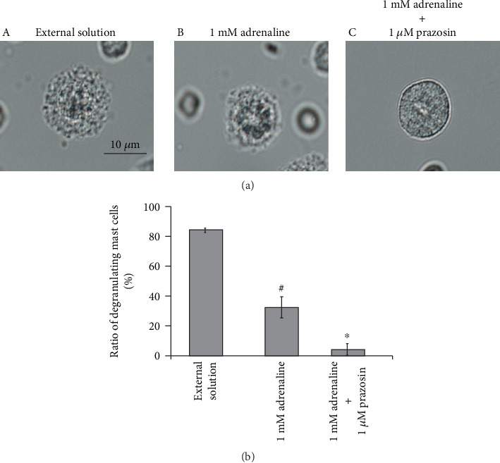
Results: Adrenaline dose-dependently suppressed the GTP-γ-S-induced increase in the Cm and inhibited the degranulation from mast cells, which was almost completely erased in the presence of butoxamine, a β 2-adrenergic receptor antagonist. Among α-adrenergic receptor agonists or antagonists, high-dose prazosin, a selective α 1-adrenergic receptor antagonist, significantly reduced the ratio of degranulating mast cells and suppressed the increase in the Cm. Additionally, prazosin augmented the inhibitory effects of adrenaline on the degranulation of mast cells.

Conclusions: This study provided electrophysiological evidence for the first time that adrenaline dose-dependently inhibited the process of exocytosis, confirming its usefulness as a potent mast cell stabilizer. The pharmacological blockade of α 1-adrenergic receptor by prazosin synergistically potentiated such mast cell-stabilizing property of adrenaline, which is primarily mediated by β 2-adrenergic receptors.


Conflict of interest statement

The authors declare no conflicts of interest.

Figures

Figure 1
Figure 1
Effects of adrenaline and dopamine on mast cell degranulation. (a) Differential-interference contrast (DIC) microscopic images were taken before (A) and after exocytosis was externally induced by compound 48/80 in mast cells incubated in the external solutions containing no drug (B), 1 μM adrenaline (C), 10 μM adrenaline (D), 100 μM adrenaline (E), 1 mM adrenaline (F), 1 μM dopamine (G), 10 μM dopamine (H), 100 μM dopamine (I), and 1 mM dopamine (J). Effects of different concentrations (1, 10, and 100 μM and 1 mM) of adrenaline (b) and dopamine (c). After the mast cells were incubated in the external solutions containing no drug or either drug, exocytosis was induced by compound 48/80. The numbers of degranulating mast cells were expressed as percentages of the total mast cell numbers in selected bright fields. #p < 0.05 vs. incubation in the external solution alone. Values are means ± SEM. Differences were analyzed by ANOVA followed by Dunnett's test.
Figure 2
Figure 2
Adrenaline-induced changes in mast cell membrane capacitance and series and membrane conductance during exocytosis. After the mast cells were incubated in the external solutions containing 1 μM (a), 10 μM (b), 100 μM (c), or 1 mM adrenaline (d), the whole-cell recording configuration was established in single mast cells and dialysis with 100 μM GTP-γ-S was started. Membrane capacitance and series and membrane conductance were monitored for at least 90 sec. Cm: membrane capacitance; Gs: series conductance; Gm: membrane conductance.
Figure 3
Figure 3
Dopamine-induced changes in mast cell membrane capacitance and series and membrane conductance during exocytosis. After the mast cells were incubated in the external solutions containing 1 μM (a), 10 μM (b), 100 μM (c), or 1 mM dopamine (d), the whole-cell recording configuration was established in single mast cells and dialysis with 100 μM GTP-γ-S was started. Membrane capacitance and series and membrane conductance were monitored for at least 90 sec. Cm: membrane capacitance; Gs: series conductance; Gm: membrane conductance.
Figure 4
Figure 4
Effects of β2-adrenergic receptor antagonist on adrenaline-induced inhibition of mast cell degranulation. (a) Differential-interference contrast (DIC) microscopic images were taken after exocytosis was externally induced by compound 48/80 in mast cells incubated in the external solutions containing no drug (A), 1 mM adrenaline (B), or 1 mM adrenaline in the presence of 1 mM butoxamine (C). (b) After exocytosis was induced in mast cells incubated in the external solutions containing no drug and 1 mM adrenaline with or without the presence of 1 mM butoxamine, the numbers of degranulating mast cells were expressed as percentages of the total mast cell numbers in selected bright fields. #p < 0.05 vs. incubation in the external solution alone. Values are means ± SEM. Differences were analyzed by ANOVA followed by Dunnett's test.
Figure 5
Figure 5
Effects of α1- or α2-adrenergic receptor agonists on mast cell degranulation. Effects of different concentrations (1, 10, and 100 μM and 1 mM) of phenylephrine (a) and clonidine (b). After the mast cells were incubated in the external solutions containing no drug or either drug, exocytosis was induced by compound 48/80. The numbers of degranulating mast cells were expressed as percentages of the total mast cell numbers in selected bright fields. Values are means ± SEM. Differences were analyzed by ANOVA followed by Dunnett's test.
Figure 6
Figure 6
Effects of α1- or α2-adrenergic receptor antagonists on mast cell degranulation. (a) Effects of prazosin on mast cell degranulation and membrane capacitance. (A) After the mast cells were incubated in the external solutions containing no drug or different concentrations (0.01, 0.1, and 1 μM) of prazosin, exocytosis was induced by compound 48/80. The numbers of degranulating mast cells were expressed as percentages of the total mast cell numbers in selected bright fields. (B) After the mast cells were incubated in the external solutions containing no drug or different concentrations (0.01, 0.1, and 1 μM) of prazosin, the whole-cell recording configuration was established in single mast cells and dialysis with 100 μM GTP-γ-S was started. The GTP-γ-S-induced increase in the Cm (⊿Cm) was calculated. (b) Effects of yohimbine on mast cell degranulation. After the mast cells were incubated in the external solutions containing no drug or different concentrations (10 and 100 μM and 1 mM) of yohimbine, exocytosis was induced by compound 48/80. The numbers of degranulating mast cells were expressed as percentages of the total mast cell numbers in selected bright fields. #p < 0.05 vs. incubation in the external solution alone. Values are means ± SEM. Differences were analyzed by ANOVA followed by Dunnett's test.
Figure 7
Figure 7
Effects of α1-adrenergic receptor antagonist on adrenaline-induced inhibition of mast cell degranulation. (a) Differential-interference contrast (DIC) microscopic images were taken after exocytosis was externally induced by compound 48/80 in mast cells incubated in the external solutions containing no drug (A), 1 mM adrenaline (B), or 1 mM adrenaline in the presence of 1 μM prazosin (C). (b) After exocytosis was induced in mast cells incubated in the external solutions containing no drug and 1 mM adrenaline with or without the presence of 1 μM prazosin, the numbers of degranulating mast cells were expressed as percentages of the total mast cell numbers in selected bright fields. #p < 0.05 vs. incubation in the external solution alone. p < 0.05 vs. incubation in the external solution containing 1 mM adrenaline. Values are means ± SEM. Differences were analyzed by ANOVA followed by Tukey's test.

Similar articles

Cited by

References

    1. Sampson H. A., Muñoz-Furlong A., Campbell R. L., et al. Second symposium on the definition and management of anaphylaxis: Summary report—Second National Institute of Allergy and Infectious Disease/Food Allergy and Anaphylaxis Network symposium. Journal of Allergy and Clinical Immunology. 2006;117(2):391–397. doi: 10.1016/j.jaci.2005.12.1303. - DOI - PubMed
    1. Kemp S. F., Lockey R. F., Simons F. E. R. Epinephrine. World Allergy Organization Journal. 2008;1(Supplement):S18–S26. doi: 10.1097/WOX.0b013e31817c9338. - DOI - PMC - PubMed
    1. Kuehn H. S., Gilfillan A. M. G protein-coupled receptors and the modification of FcepsilonRI-mediated mast cell activation. Immunology Letters. 2007;113(2):59–69. doi: 10.1016/j.imlet.2007.08.007. - DOI - PMC - PubMed
    1. Schulze W., Fu M. L. Localization of alpha 1-adrenoceptors in rat and human hearts by immunocytochemistry. Molecular and Cellular Biochemistry. 1996;163-164:159–165. doi: 10.1007/bf00408653. - DOI - PubMed
    1. Kaliner M., Orange R. P., Austen K. F. Immunological release of histamine and slow reacting substance of anaphylaxis from human lung. The Journal of Experimental Medicine. 1972;136(3):556–567. doi: 10.1084/jem.136.3.556. - DOI - PMC - PubMed

Publication types

LinkOut - more resources