

Insights into Substrate and Inhibitor Selectivity among Human GLUT Transporters through Comparative Modeling and Molecular Docking
- PMID: 32462103
- PMCID: PMC7244221
- DOI: 10.1021/acsomega.8b03447
Insights into Substrate and Inhibitor Selectivity among Human GLUT Transporters through Comparative Modeling and Molecular Docking
Abstract
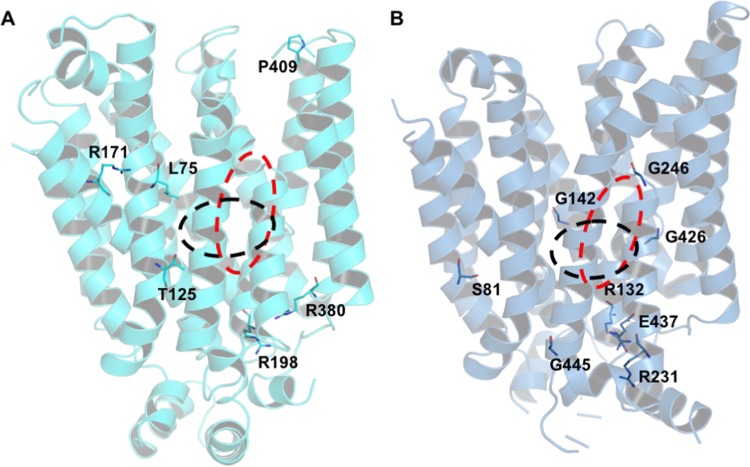
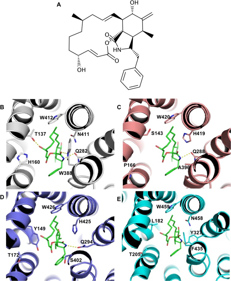
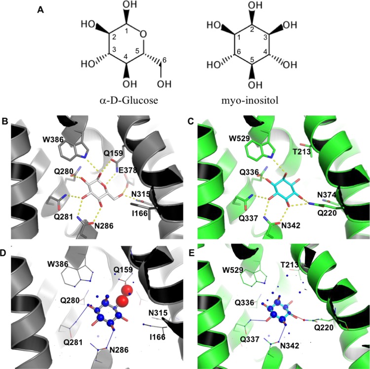
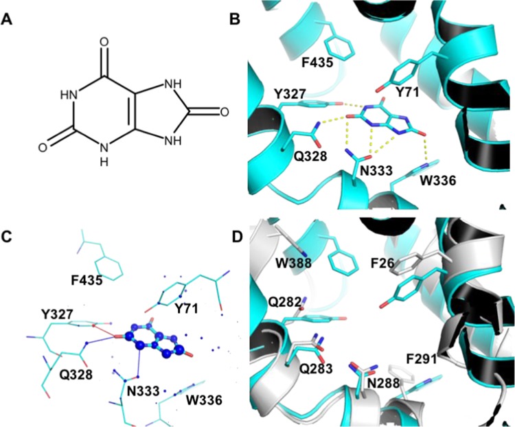
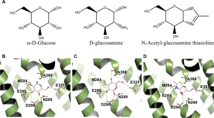
The solute carrier 2 family is composed of 14 transporters, which are members of the major facilitator superfamily. Despite their high physiological importance, there are still many open questions concerning their function and specificity, and in some cases, their physiological substrate is still unknown. To understand the determinants of the substrate and inhibitor specificity, we modeled all human glucose transport carriers (GLUTs) and simulated their interaction with known ligands. Comparative modeling was performed with the @TOME-2 pipeline, employing multiple templates and providing an ensemble of models for each GLUT. We analyzed models in both outward-occluded and inward-open conformations, to compare exofacial and endofacial binding sites throughout the family and understand differences in susceptibility of GLUTs to the inhibitor cytochalasin B. Finally, we employed molecular docking and bioinformatics to identify residues likely critical for recognition of myo-inositol by GLUT13 and urate by GLUT9. These results provide insights into the molecular basis for the specificity for these substrates. In addition, we suggested a potential recognition site of glucosamine by GLUT11 to be evaluated in future experiments.
Copyright © 2019 American Chemical Society.
Conflict of interest statement
The authors declare no competing financial interest.
Figures
Similar articles
-
Molecular basis of ligand recognition and transport by glucose transporters.Nature. 2015 Oct 15;526(7573):391-6. doi: 10.1038/nature14655. Epub 2015 Jul 15. Nature. 2015. PMID: 26176916
-
The extended GLUT-family of sugar/polyol transport facilitators: nomenclature, sequence characteristics, and potential function of its novel members (review).Mol Membr Biol. 2001 Oct-Dec;18(4):247-56. doi: 10.1080/09687680110090456. Mol Membr Biol. 2001. PMID: 11780753 Review.
-
Characterization of human glucose transporter (GLUT) 11 (encoded by SLC2A11), a novel sugar-transport facilitator specifically expressed in heart and skeletal muscle.Biochem J. 2001 Oct 15;359(Pt 2):443-9. doi: 10.1042/0264-6021:3590443. Biochem J. 2001. PMID: 11583593 Free PMC article.
-
A Glimpse of Membrane Transport through Structures-Advances in the Structural Biology of the GLUT Glucose Transporters.J Mol Biol. 2017 Aug 18;429(17):2710-2725. doi: 10.1016/j.jmb.2017.07.009. Epub 2017 Jul 26. J Mol Biol. 2017. PMID: 28756087 Review.
-
Avian and Mammalian Facilitative Glucose Transporters.Microarrays (Basel). 2017 Apr 5;6(2):7. doi: 10.3390/microarrays6020007. Microarrays (Basel). 2017. PMID: 28379195 Free PMC article. Review.
Cited by
-
Identification of Structural Determinants of the Transport of the Dehydroascorbic Acid Mediated by Glucose Transport GLUT1.Molecules. 2023 Jan 5;28(2):521. doi: 10.3390/molecules28020521. Molecules. 2023. PMID: 36677580 Free PMC article.
-
Therapeutic effects and mechanisms of N-(9,10-anthraquinone-2-ylcarbonyl) xanthine oxidase inhibitors on hyperuricemia.Front Pharmacol. 2022 Sep 2;13:950699. doi: 10.3389/fphar.2022.950699. eCollection 2022. Front Pharmacol. 2022. PMID: 36120294 Free PMC article.
-
Towards Selective Binding to the GLUT5 Transporter: Synthesis, Molecular Dynamics and In Vitro Evaluation of Novel C-3-Modified 2,5-Anhydro-D-mannitol Analogs.Pharmaceutics. 2022 Apr 10;14(4):828. doi: 10.3390/pharmaceutics14040828. Pharmaceutics. 2022. PMID: 35456662 Free PMC article.
-
Progress of research on glucose transporter proteins in hepatocellular carcinoma.World J Hepatol. 2025 Mar 27;17(3):104715. doi: 10.4254/wjh.v17.i3.104715. World J Hepatol. 2025. PMID: 40177207 Free PMC article. Review.
-
Identification of druggable small molecule antagonists of the Plasmodium falciparum hexose transporter PfHT and assessment of ligand access to the glucose permeation pathway via FLAG-mediated protein engineering.PLoS One. 2019 May 9;14(5):e0216457. doi: 10.1371/journal.pone.0216457. eCollection 2019. PLoS One. 2019. PMID: 31071153 Free PMC article.
References
LinkOut - more resources
Full Text Sources