. 2020 Jul;104(14):6173-6183.
doi: 10.1007/s00253-020-10691-z. Epub 2020 May 27.

Production and characterization of a monoclonal antibody against the sialidase of Gardnerella vaginalis using a synthetic peptide in a MAP8 format

Affiliations

Production and characterization of a monoclonal antibody against the sialidase of Gardnerella vaginalis using a synthetic peptide in a MAP8 format

Karen Cortés-Sarabia et al. Appl Microbiol Biotechnol. 2020 Jul.

Abstract

Bacterial vaginosis is one of the most frequent vaginal infections. Its main etiological agent is Gardnerella vaginalis, which produces several virulence factors involved in vaginal infection and colonization, in particular, sialidase (SLD), a potential clinical biomarker that participates in immune response modulation and mucus degradation. The main objective of this work was the production and evaluation of a monoclonal antibody against G. vaginalis sialidase and its validation in immunoassays. For immunization of mice, a synthetic multiantigenic peptide was used, and hybridomas were generated. After fusion, hybridomas were evaluated for antibody production and cloned by limited dilution. One clone producing IgG1 was selected and characterized by indirect ELISA, dot blot, and Western blot, and we also tested clinical isolates and HeLa cells infected with G. vaginalis. The results showed that the anti-SLD antibody recognized a single protein of ~90 kDa that correlated with the estimated molecular weight of SLD. In addition, anti-SLD antibody recognized SLD from complete bacteria and from culture supernatants of infected Hela cells. In conclusion, our results showed that the anti-SLD antibody recognized SLD from different sources and could be considered a new tool for the diagnosis of bacterial vaginosis. KEY POINTS: • Anti-sialidase mAb was generated using a synthetic peptide • The mAb recognizes synthetic peptide and intact protein from multiple sources • The antibody was characterized by several immunological methods.

Keywords: Bacterial vaginosis; Gardnerella vaginalis; Monoclonal antibody; Sialidase.


Conflict of interest statement

The authors declare that they have no conflict of interest.

Figures

Fig. 1
Fig. 1
Immune response evaluation and anti-SLD clones. (a) Immune response evaluation in selected mice. Indirect ELISA using as primary antibody the collected serum during the immunization process. As a secondary antibody, we used an anti-H + L antibody. (b) Evaluation of clones obtained after fusion. Indirect ELISA using as primary antibody the culture supernatant of the evaluated clones in order to determine the immunoglobulin class produced by each clone. We used specific antibodies against the mu and gamma chains. (c) and (d) Representative images of the hybridomas produced
Fig. 2
Fig. 2
Anti-SLD mAb. (a) Immunoglobulin class and subclass produced by the 1H12 clone. As primary antibody, we used the culture supernatant of clone 1H12, mouse hyperimmune serum (C+), and no hyperimmune serum (C−). As secondary antibodies, we used specific antibodies against different classes (IgM and IgG) and subclasses (IgG1, IgG2a, IgG2b, and IgG3). (b) Anti-SLD mAb purification evaluation. 12% SDS-PAGE and Coomassie blue staining. MWM, molecular weight marker; HC, heavy chain; LC, light chain. (c) and (d) Antibody and antigen titration. Indirect ELISA using decreasing concentrations of antigen and purified antibody by double dilution. As primary antibody, we used purified 1H12 antibody, and as secondary antibody, an anti-H + L antibody was used
Fig. 3
Fig. 3
Preliminary characterization of 1H12 anti-SLD mAb. (a) and (b) Western blot and dot blot analyses. We used total lysates, complete bacteria, or culture supernatants of G. vaginalis ATCC 14018 strain and clinical isolates of biotypes 1, 2, 5, and 6 as antigens. (c) and (d) Detection of SLD in the ATCC 14018 strain and clinically isolated biotypes of G. vaginalis. Indirect ELISA using protein total extract and proteins precipitated from the culture supernatants to evaluate antibody recognition. In all experiments, 1H12 anti-SLD mAb, C+ is polyclonal antibody against G. vaginalis and in C− primary antibody was omitted. As secondary antibody, an anti-H + L antibody was used
Fig. 4
Fig. 4
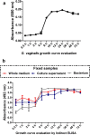
Evaluation of SLD recognition during growth of G. vaginalis (a) Growth curve of G. vaginalis. We used as culture medium brain heart infusion, and optical density was determined at 600 nm during 0.5, 1, 2, 4, 8, 10, 12, 24, 36, 48, and 72 h. (b) Detection of SLD during the growth of G. vaginalis. Indirect ELISA for qualitative evaluation of the SLD present in samples of complete bacterial culture (bacteria + culture supernatant), culture supernatant, or bacteria only. As primary antibody, we used the 1H12 anti-SLD antibody and as secondary antibody, we used an anti-H + L antibody
Fig. 5
Fig. 5
SLD recognition in clinical samples. (a) and (b) Dot blot results of SLD evaluation in the complete bacteria and culture supernatants. Each dot is a different clinical sample, and we used the same sample in the same position in the complete bacteria and culture supernatants. Dot blots were performed as previously described. Negative controls: E8–9, H7–9; positive controls: E10, H10. (c) and (d) Densitometric analysis of the dot blots. The analysis was carried out using ImageJ software
Fig. 6
Fig. 6
Evaluation of SLD in HeLa cells infected with G. vaginalis (a) Representative images of HeLa cells infected with G. vaginalis. Cells were infected with a MOI (multiplicity of infection) of 1 and 20 over 2 and 4 h. (b) Indirect ELISA and (c) Dot blot evaluation of SLD in infected HeLa cells. HPI, hours post-infection. The primary antibody used was 1H12, and as a secondary antibody, we used an anti-H + L antibody

Similar articles

Cited by

References

    1. Amsel R, Totten PA, Spiegel CA, Chen KC, Eschenbach D, Holmes KK. Nonspecific vaginitis. Diagnostic criteria and microbial and epidemiologic associations. Am J Med. 1983;74:14–22. doi: 10.1016/0002-9343(83)91112-9. - DOI - PubMed
    1. Bautista CT, Wurapa E, Sateren WB, Morris S, Hollingsworth B, Sanchez JL. Bacterial vaginosis: a synthesis of the literature on etiology, prevalence, risk factors, and relationship with chlamydia and gonorrhea infections. Mil Med Res. 2016;3:1–10. doi: 10.1186/s40779-016-0074-5. - DOI - PMC - PubMed
    1. Bordeaux J, Welsh A, Agarwal S, Killiam E, Baquero M, Hanna J, Anagnostou V, Rimm D. Antibody validation. BioTechniques. 2010;48:197–209. doi: 10.2144/000113382. - DOI - PMC - PubMed
    1. Briselden AM, Moncla BJ, Stevens CE, Hillier SL. Sialidases (neuraminidases) in bacterial vaginosis and bacterial vaginosis-associated microflora. J Clin Microbiol. 1992;30:663–666. doi: 10.1128/JCM.30.3.663-666.1992. - DOI - PMC - PubMed
    1. Buve A, Jespers V, Crucitti T, Fichorova RN. The vaginal microbiota and susceptibility to HIV. AIDS Lond Engl. 2014;28:2333–2344. doi: 10.1097/qad.0000000000000432. - DOI - PubMed

MeSH terms

Grants and funding

LinkOut - more resources