

Lymphoepithelial Carcinoma of Salivary Gland EBV-association in Endemic versus Non-Endemic Patients: A Report of 16 Cases
- PMID: 32462279
- PMCID: PMC7669917
- DOI: 10.1007/s12105-020-01172-w
Lymphoepithelial Carcinoma of Salivary Gland EBV-association in Endemic versus Non-Endemic Patients: A Report of 16 Cases
Abstract
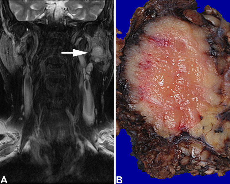
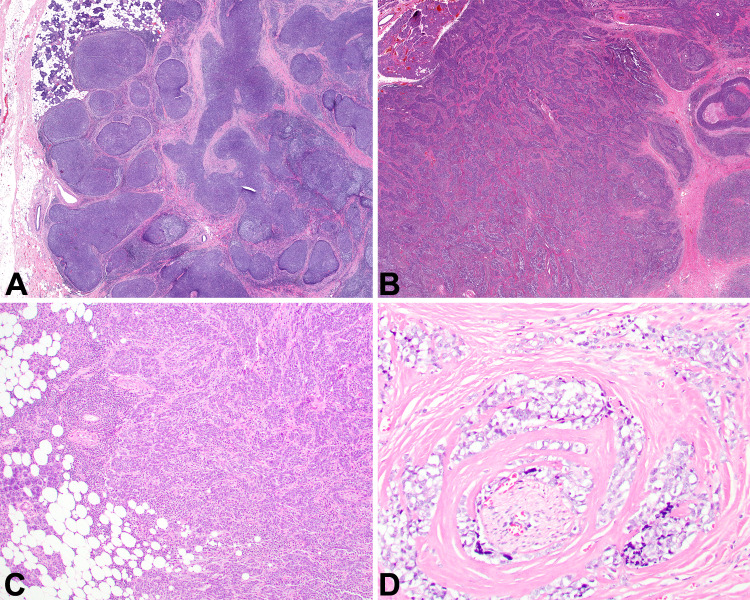
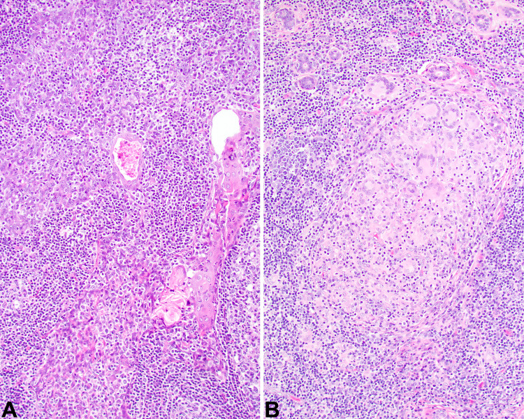
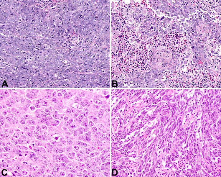
Lymphoepithelial carcinoma of salivary glands (LECSG) are rare neoplasms, reported in endemic populations (southeastern Chinese) with a strong Epstein-Barr virus (EBV) association. A retrospective series comparing EBV status within an ethnically diverse population (endemic vs. non-endemic patients) has not been reported. Sixteen LECSG were equally distributed between males (n = 8) and females (n = 8) with a median age of 54 years (range 18 to 85 years) at initial diagnosis. Ten patients were white, 4 Asian, and 2 black. The patients typically presented with swelling or mass for an average of 11.6 months. Tumors affected only major salivary glands: parotid (n = 13); submandibular (n = 3). Tumors were an average of 2.9 cm (range 1.5 to 5.8 cm). Nine of 16 (56%) patients had cervical lymph node metastases at presentation. No patients had nasopharyngeal or oropharyngeal tumors. Microscopically, the tumors were widely infiltrative, characterized by large polygonal to spindled cells arranged in a syncytial, lattice-like network in a background of lymphoplasmacytic cells. The neoplastic cells showed an open-vesicular nuclear chromatin to a more basaloid-morphology, the latter showing hyperchromatic nuclei and less cytoplasm, while nearly all of the cases had associated lymphoepithelial lesions/sialadenitis. By in situ hybridization, 8 of 16 cases had a strong, diffuse EBER expression (4 of 4 Asians; 4 of 12 non-Asians), while with immunohistochemistry all cases tested were pan-cytokeratin, CK5/6 and p63 reactive; none of the cases tested were p16 reactive. All patients were managed with wide or radical excision, 4 with concurrent chemoradiation, and 6 with radiation alone. Distant metastasis (lung, brain, and bone) developed in 2 patients. Overall follow-up (mean 3.8 years) revealed 12 patients alive and 2 dead, none with evidence of disease (mean 4.3 years); one white male alive with disease at 1.9 years, and one Asian female dead of disease at 4.2 years; both of these latter patients had Group IV stage disease. High stage (Group IV) patients had a shorter mean survival than lower stage patients: 3.1 versus 4.8 years, respectively. In conclusion, LECSG are uncommon primary neoplasms. Concurrent lymphoepithelial lesions may help suggest a primary tumor. The tumors, irrespective of race or ethnicity, may express EBER. There is an overall good survival, perhaps better for EBV-negative patients and for those with lower stage disease.
Keywords: EBER; Ethnic groups; Immunohistochemistry; Lymphoepithelial carcinoma; Salivary glands.
Conflict of interest statement
All authors declare that they have no conflict of interest as it relates to this research project.
Figures


References
-
- Lewis JS, El-Mofty SK, Nicolai P. Lymphoepithelial Carcinoma. In: El-Naggar AK, Chan JKC, Grandis JR, Takata T, Slootweg PJ, editors. WHO classification of head and neck tumors. Lyon: IARC; 2017. pp. 181–182.
-
- Cleary KR, Batsakis JG. Undifferentiated carcinoma with lymphoid stroma of the major salivary glands. Ann Otol Rhinol Laryngol. 1990;99:236–238. - PubMed
-
- Thompson LDR. Head and neck cancer. In: Stewart BW, Wild CP, editors. World cancer report. Lyon: IARCPress; 2014. pp. 422–435.
-
- Wenig BM. Lymphoepithelial-like carcinomas of the head and neck. Semin Diagn Pathol. 2015;32:74–86. - PubMed
MeSH terms
LinkOut - more resources
Full Text Sources
Medical
Research Materials
