. 2020 Dec;267(Suppl 1):126-135.
doi: 10.1007/s00415-020-09909-x. Epub 2020 May 27.

Risk of acute brain lesions in dizzy patients presenting to the emergency room: who needs imaging and who does not?

Affiliations

Risk of acute brain lesions in dizzy patients presenting to the emergency room: who needs imaging and who does not?

Björn Machner et al. J Neurol. 2020 Dec.

Abstract

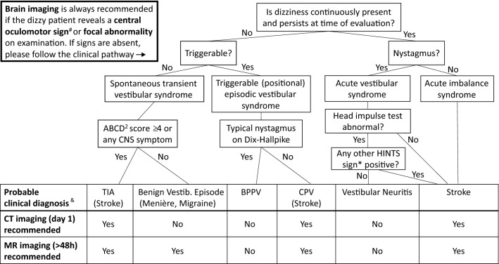
The usefulness of brain imaging studies in dizzy patients presenting to the emergency department (ED) is controversial. We aimed to assess the 'real-world' probability of ischemic stroke and other acute brain lesions (ABLs) in these patients to create an algorithm that helps decision-making on whether which and when brain imaging is needed. By reviewing medical records, we identified 610 patients presenting with dizziness, vertigo or imbalance to our university hospital's ED and receiving neurological workup. We collected timing/triggers of symptoms, ABCD2 score, focal neurological abnormalities, HINTS (head impulse, nystagmus, test-of-skew) and other central oculomotor signs. ABLs were extracted from CT/MRI reports. Uni-/multivariate logistic regression analyses investigated associations between clinical parameters and ABLs. Finally, the likelihood of ABLs was assessed for different clinically defined subgroups ('dizziness syndromes'). Early CT (day 1) was performed in 539 (88%) and delayed MR imaging (median: day 4) in 299 (49%) patients. ABLs (89% ischemic stroke) were revealed in 75 (24%) of 318 patients with adequate imaging (MRI or lesion-positive CT). The risk for ABLs increased with the presence of central oculomotor signs (odds ratio 2.8, 95% confidence interval 1.5-5.2) or focal abnormalities (OR 3.3, 95% CI 1.8-6.2). The likelihood of ABLs differed between dizziness syndromes, e.g., HINTS-negative acute vestibular syndrome: 0%, acute imbalance syndrome with ABCD2-score ≥ 4: 50%. We propose a clinical pathway, according to which patients with HINTS-negative acute vestibular syndrome should not receive brain imaging, whereas imaging is suggested in dizzy patients with acute imbalance, central oculomotor signs or focal abnormalities.

Keywords: CT; Dizziness; MRI; Nystagmus; Stroke; Vertigo.


Conflict of interest statement

The authors declare that they have no conflicts of interest.

Figures

Fig. 1
Fig. 1
Probability of an acute brain lesion as detected by early CT a or delayed MR imaging b in dependence of the patients’ clinical sub-specification (‘dizziness syndrome’). For the purpose of clarity, we color coded the probability/risk to have an acute brain lesion (ABL) revealed by the respective imaging study (green: no risk, yellow: low–medium risk, red: high risk). a Early CT imaging performed in 534 of 610 ‘dizzy’ patients revealed ABLs in 36 of them (5.9%). Further stratification of the patients by using information from targeted history taking and clinical examination can increase the probability of detecting ABLs on CT to over 20% (e.g., ‘acute imbalance syndrome (AIS) with high-risk ABCD2-score’). b Delayed MRI is more sensitive in detecting ABLs and identifies high-risk subgroups (e.g., ABLs in 50% of AIS patients with ABCD2 ≥ 4), but also no-risk subgroups (e.g., 0% ABLs in HINTS-negative AVS patients). Notably, b also includes those patients with an ABL already detected on the early CT (‘lesion-positive’ CT) who did not receive a redundant MRI. *Patients with benign paroxysmal positional vertigo (BPPV) were generally rare in our study cohort as they were usually identified and directly discharged from the ED
Fig. 2
Fig. 2
Clinical pathway to help decision-making on brain imaging in patients presenting to the ED with dizziness, vertigo or imbalance and without a general medical cause+. TIA transient ischemic attack, BPPV benign paroxysmal positional vertigo, CPV central positional vertigo. +General medical causes comprise various toxic, metabolic, infectious, or cardiovascular diseases (see Edlow et al. 2018 [8]). #Central oculomotor signs include: vertical or purely torsional spontaneous nystagmus, horizontal/vertical gaze-evoked nystagmus, gaze palsies, bilaterally disrupted smooth pursuit eye movements.*HINTS are positive (‘central’) if any of the following signs is present: normal head impulse test, the nystagmus’ fast phase alternating with gaze, skew deviation with a refixation on cover test. §The MRI may be dispensable if the lesion has already been detected by early CT. &Only the most likely and most relevant differential diagnosis is stated.

References

    1. Brazzelli M, Chappell FM, Miranda H, Shuler K, Dennis M, Sandercock PA, Muir K, Wardlaw JM. Diffusion-weighted imaging and diagnosis of transient ischemic attack. Ann Neurol. 2014;75:67–76. doi: 10.1002/ana.24026. - DOI - PMC - PubMed
    1. Chalela JA, Kidwell CS, Nentwich LM, Luby M, Butman JA, Demchuk AM, Hill MD, Patronas N, Latour L, Warach S. Magnetic resonance imaging and computed tomography in emergency assessment of patients with suspected acute stroke: a prospective comparison. Lancet. 2007;369:293–298. doi: 10.1016/S0140-6736(07)60151-2. - DOI - PMC - PubMed
    1. Choi JH, Park MG, Choi SY, Park KP, Baik SK, Kim JS, Choi KD. Acute transient vestibular syndrome: prevalence of stroke and efficacy of bedside evaluation. Stroke. 2017;48:556–562. doi: 10.1161/STROKEAHA.116.015507. - DOI - PubMed
    1. Choi SY, Kee HJ, Park JH, Kim HJ, Kim JS. Combined peripheral and central vestibulopathy. J Vestib Res. 2014;24:443–451. doi: 10.3233/VES-140524. - DOI - PubMed
    1. Cnyrim CD, Newman-Toker D, Karch C, Brandt T, Strupp M. Bedside differentiation of vestibular neuritis from central “vestibular pseudoneuritis”. J Neurol Neurosurg Psychiatr. 2008;79:458–460. doi: 10.1136/jnnp.2007.123596. - DOI - PubMed