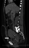
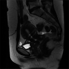
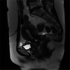
Magnetic resonance imaging of the female pelvis after Cesarean section: a pictorial review
- PMID: 32462368
- PMCID: PMC7253593
- DOI: 10.1186/s13244-020-00876-5
Magnetic resonance imaging of the female pelvis after Cesarean section: a pictorial review
Abstract
The rate of Cesarean sections (C-sections) in Poland increased from 21.7% in 2001 to 43.85% in 2017 even though the Polish Society of Gynecologists and Obstetricians highlights the negative consequences of C-section for both mother and child and recommends to make every possible effort to reduce its percentage, following the World Health Organization recommendations. There is a long list of possible complications related to the uterine scar after C-section, including uterine scar dehiscence, uterine rupture, abdominal and pelvic adhesions, uterine synechiae, ectopic pregnancy, anomalous location of the placenta, placental invasion, and-rarely-vesicouterine or uterocutaneous fistulas. Ultrasound (US) remains the first-line modality; however, its strong operator- and equipment dependence and other limitations require further investigations in some cases. Magnetic resonance imaging (MRI) is the second-line tool which is supposed to confirm, correct, or complete the sonographic diagnosis thanks to its higher tissue resolution and bigger field of view. This article will discuss the spectrum of C-section complications in the MR image-rich form and will provide a systematic discussion of the possible pathology that can occur, showing comprehensive anatomical insight into the pelvis after C-section thanks to MRI that facilitates clinical decisions.
Keywords: Cesarean section (C-section); Magnetic resonance imaging (MRI); Pelvis.
Conflict of interest statement
The author declares that she has no competing interests.
Figures


Similar articles
-
Cesarean Scar Ectopic Pregnancy: Laparoscopic Resection and Total Scar Dehiscence Repair.J Minim Invasive Gynecol. 2018 Feb;25(2):297-298. doi: 10.1016/j.jmig.2017.01.022. Epub 2017 Feb 4. J Minim Invasive Gynecol. 2018. PMID: 28179198
-
Vesicouterine fistulas following cesarean section: report on a case, review and update of the literature.Int Urol Nephrol. 2002;34(3):335-44. doi: 10.1023/a:1024443822378. Int Urol Nephrol. 2002. PMID: 12899224 Review.
-
Robotic-assisted Laparoscopic Repair of a Cesarean Section Scar Defect.J Minim Invasive Gynecol. 2015 Nov-Dec;22(7):1135-6. doi: 10.1016/j.jmig.2015.06.001. Epub 2015 Jun 10. J Minim Invasive Gynecol. 2015. PMID: 26070729
-
Laparoscopic Resection of Cesarean Scar Ectopic Pregnancy after Unsuccessful Systemic Methotrexate Treatment.J Minim Invasive Gynecol. 2019 Mar-Apr;26(3):399-400. doi: 10.1016/j.jmig.2018.06.003. Epub 2018 Jun 8. J Minim Invasive Gynecol. 2019. PMID: 29890356
-
Delivery for women with a previous cesarean: guidelines for clinical practice from the French College of Gynecologists and Obstetricians (CNGOF).Eur J Obstet Gynecol Reprod Biol. 2013 Sep;170(1):25-32. doi: 10.1016/j.ejogrb.2013.05.015. Epub 2013 Jun 28. Eur J Obstet Gynecol Reprod Biol. 2013. PMID: 23810846 Review.
Cited by
-
Anterior uterine incarceration complicated by placenta previa and placenta accreta spectrum disorder: a case report.Ann Transl Med. 2023 Jan 31;11(2):139. doi: 10.21037/atm-22-5158. Epub 2023 Jan 3. Ann Transl Med. 2023. PMID: 36819576 Free PMC article.
-
Endometriosis MR mimickers: T2-hypointense lesions.Insights Imaging. 2024 Jan 25;15(1):20. doi: 10.1186/s13244-023-01588-2. Insights Imaging. 2024. PMID: 38267633 Free PMC article. Review.
References
-
- World Health Organization Human Reproduction Programme (2015) WHO Statement on caesarean section rates. Reprod Health Matters. 23(45):149–150. 10.1016/j.rhm.2015.07.007 - PubMed
-
- Orhan Adnan, Kasapoğlu Işıl, Çetinkaya Demir Bilge, Özerkan Kemal, Duzok Nergis, Uncu Gürkan. Different treatment modalities and outcomes in cesarean scar pregnancy: a retrospective analysis of 31 cases in a unıversity hospital. Ginekologia Polska. 2019;90(6):291–307. doi: 10.5603/GP.2019.0053. - DOI - PubMed
-
- OECD (2019) Caesarean sections (indicator). doi: 10.1787/adc3c39f-en. Accessed 15 Dec 2019.
Publication types
LinkOut - more resources
Full Text Sources

