Any unique image biomarkers associated with COVID-19?
- PMID: 32462445
- PMCID: PMC7253230
- DOI: 10.1007/s00330-020-06956-w
Any unique image biomarkers associated with COVID-19?
Abstract
Objective: To define the uniqueness of chest CT infiltrative features associated with COVID-19 image characteristics as potential diagnostic biomarkers.
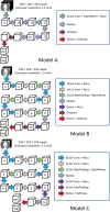
Methods: We retrospectively collected chest CT exams including n = 498 on 151 unique patients RT-PCR positive for COVID-19 and n = 497 unique patients with community-acquired pneumonia (CAP). Both COVID-19 and CAP image sets were partitioned into three groups for training, validation, and testing respectively. In an attempt to discriminate COVID-19 from CAP, we developed several classifiers based on three-dimensional (3D) convolutional neural networks (CNNs). We also asked two experienced radiologists to visually interpret the testing set and discriminate COVID-19 from CAP. The classification performance of the computer algorithms and the radiologists was assessed using the receiver operating characteristic (ROC) analysis, and the nonparametric approaches with multiplicity adjustments when necessary.
Results: One of the considered models showed non-trivial, but moderate diagnostic ability overall (AUC of 0.70 with 99% CI 0.56-0.85). This model allowed for the identification of 8-50% of CAP patients with only 2% of COVID-19 patients.
Conclusions: Professional or automated interpretation of CT exams has a moderately low ability to distinguish between COVID-19 and CAP cases. However, the automated image analysis is promising for targeted decision-making due to being able to accurately identify a sizable subsect of non-COVID-19 cases.
Key points: • Both human experts and artificial intelligent models were used to classify the CT scans. • ROC analysis and the nonparametric approaches were used to analyze the performance of the radiologists and computer algorithms. • Unique image features or patterns may not exist for reliably distinguishing all COVID-19 from CAP; however, there may be imaging markers that can identify a sizable subset of non-COVID-19 cases.
Keywords: Biomarkers; COVID-19; Neural network; Pneumonia.
Conflict of interest statement
The authors declare that there is no conflict of interest.
Figures


References
-
- Ufuk F (2020) 3D CT of novel coronavirus (COVID-19) pneumonia. Radiology. 201183. 10.1148/radiol.2020201183
MeSH terms
Substances
Grants and funding
LinkOut - more resources
Full Text Sources
Medical
Miscellaneous

