Review
. 2020 May 27;11(1):74.
doi: 10.1186/s13244-020-00867-6.

Testicular tumours in children: an approach to diagnosis and management with pathologic correlation

Affiliations
Review

Testicular tumours in children: an approach to diagnosis and management with pathologic correlation

Cinta Sangüesa et al. Insights Imaging. .

Abstract

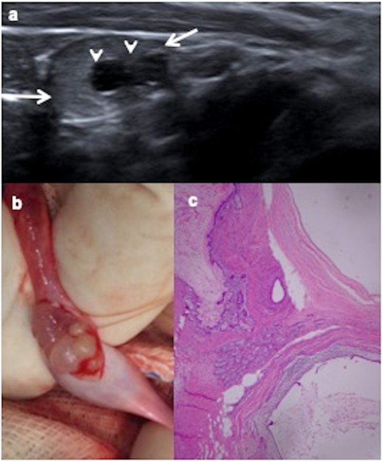
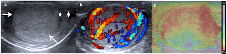
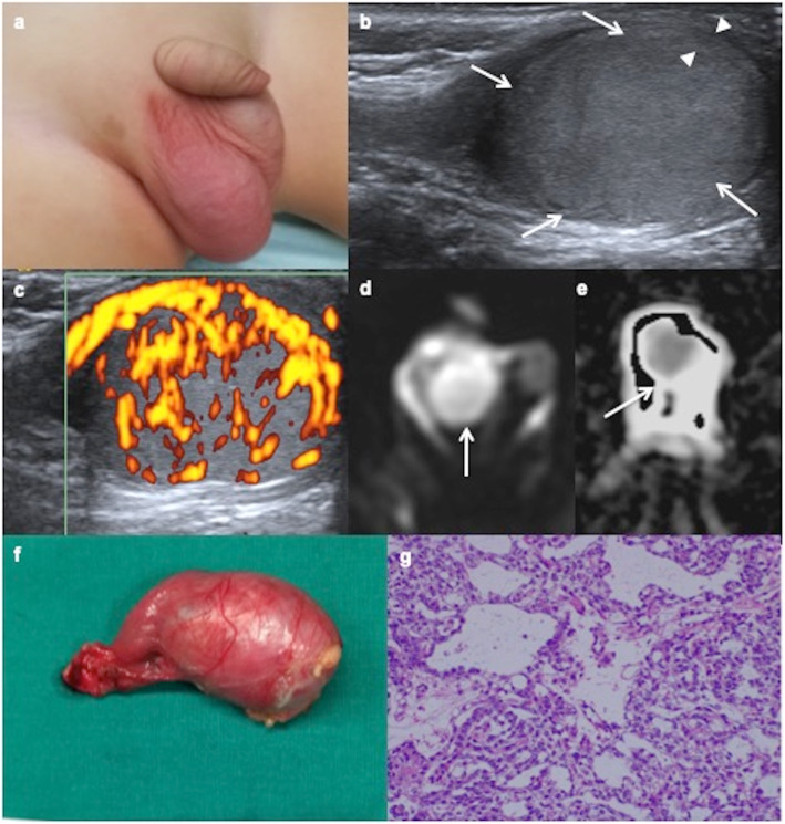
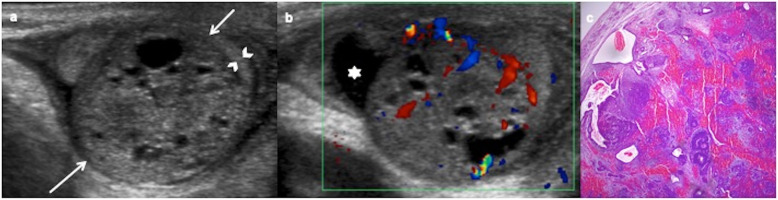
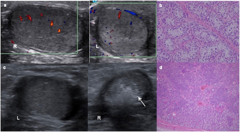
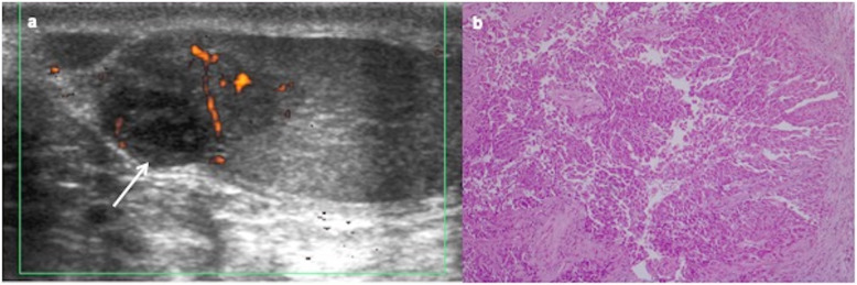
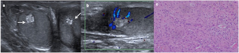
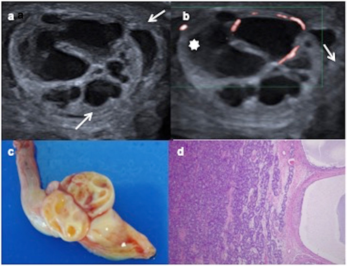
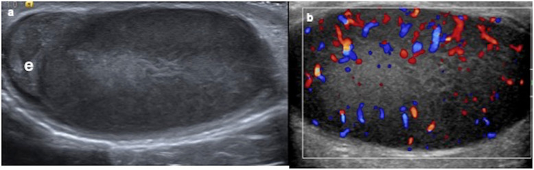
Testicular tumours are rare in children. Painless scrotal mass is the most frequent clinical presentation. Tumoural markers (alpha-fetoprotein, beta-human gonadotropin chorionic) and hormone levels (testosterone) contribute to the diagnosis and management of a testicular mass in boys. Ultrasonography is the best imaging modality to study testicular tumours. A benign tumour is suggested when ultrasonography shows a mainly cystic component, well-defined borders, echogenic rim or normal to increased echogenicity lesion when compared to the healthy testicular parenchyma. Malignant tumour is suspected when ultrasonography shows inhomogeneous, hypoechoic, not well-circumscribed or diffuse infiltration lesion. However, these ultrasonographic findings may overlap. Colour Doppler, power Doppler, elastography and contrast-enhanced ultrasonography are useful complementary methods to characterise the focal testicular lesions. Chest computerised tomography and abdominopelvic magnetic resonance are necessary to establish the extension in case of malignant proved tumours.Benign tumours are more frequent in prepuberal boys and malignant tumours in pubertal boys. Mature teratoma prepubertal-type is the most common histologic type. Testicular sparing surgery is the choice in benign tumours. Radical inguinal orchiectomy is indicated in malignant tumours. Prognostic is excellent.The purpose of our study is to show an approach to the diagnosis and management of the most frequent testicular tumours in children according to clinical manifestations, imaging findings and tumour markers levels based on histologically confirmed tumours in our hospital.

Keywords: Alpha-fetoprotein; Child; Testicular neoplasm; Ultrasonography, Doppler.


Conflict of interest statement

The authors declare that they have no competing interests.

Figures

Fig. 1
Fig. 1
Cystic mature teratoma in cryptorchidism. A 10-month-old boy with right cryptorchidism. a Sonography identifies the right testis (arrows) in the inguinal canal presenting two cystic lesions (arrowheads). b Surgical procedure shows the testis during tumourectomy where cystic lesions are visible. c Cystic mature teratoma is the definitive histopathologic result. Microscopic view of the tumour shows squamous, digestive and ciliated columnar epithelium. No immature elements are identified
Fig. 2
Fig. 2
Tumour and microlithiasis. A 12-year-old boy with right scrotal pain. a Longitudinal ultrasound view of the left testicle shows multiple punctuate non-shadowing echogenicities compatible with microlithiasis. b Longitudinal view of the right testicle shows besides microlithiasis, a round well-defined lesion with onion ring appearance (arrow) typically characteristic of epidermoid cyst. c Colour Doppler shows only peripheral vascularization. An intraoperative biopsy diagnoses an epidermoid cyst and a testis sparing surgery is practised
Fig. 3
Fig. 3
Yolk sac tumour. A 15-month-old boy with a painless swollen left testicle. a Longitudinal view of the left testis shows near-complete replacement by a hypoechoic solid mass (arrows) with a rim of normal parenchyma (arrowheads). b Power Doppler reveals increased blood flow within the tumour. c Elastography shows heterogeneous with high strain of the mass. The boy has a very high level of alphaphetoprotein and inguinal orchiectomy is practised. Yolk sac tumour is the definitive pathologic diagnosis
Fig. 4
Fig. 4
Yolk sac tumour. a A 14-month-old boy with a painless right scrotal mass. b Ultrasound shows an intratesticular hypoechoic mass (arrows) with a rim of healthy tissue preserved (arrowheads). c Power Doppler reveals abnormally abundant blood flow within the tumour. d Axial diffusion-weighted MRI shows the mass with hyperintensity (arrow). e ADC map shows a low signal intensity from diffusion restriction of the mass (arrow). The boy has a very high level of alphaphetoprotein and inguinal orchiectomy is practised. f Surgical piece includes inguinal cord. g Microscopic exam shows a microcystic yolk sac tumour containing glands and tubular structures with subnuclear vacuoles.
Fig. 5
Fig. 5
Yolk sac tumour. A 19-month-old boy with a painless swollen right testicle. High level of alphaphetoprotein (3.450 ng/mL). a Longitudinal view of the right testis shows near-complete replacement by a hypoechoic solid mass (arrows) with a rim of normal parenchyma (arrowheads). b Power Doppler reveals increased blood flow within the tumour. c, d Axial fat-supressed T1 show a hypointense mass with avid enhancement of the tumour after the contrast. e Surgical piece after the radical inguinal orchiectomy
Fig. 6
Fig. 6
Mature teratomas, prepubertal-type: ultrasound appearances. a, b Ultrasound and colour Doppler of a 10-month-old boy with a swollen and tender left testis show an avascular cystic mass completely replacing the testicular parenchyma, an orchiectomy being required. Right testis (discontinuous arrow). c, d Ultrasound and colour Doppler of a 8-month-old boy with a painless scrotal left mass show a mixed solid-cystic mass (arrows) without blood flow in the solid part of the lesion. Orchiectomy is practised because of the big size of the lesion. e, f Ultrasound and colour Doppler of a 8-year-old boy with a swollen and hard right testis show a heterogeneous solid mass, calcifications (arrow) and blood flow inside. A testis sparing surgery is practised since sufficient salvage parenchyma is visible
Fig. 7
Fig. 7
Immature teratoma, prepubertal-type. A 31-day-old boy with a painless left scrotal mass. a Ultrasound shows a heterogeneous predominantly solid mass (arrows) replacing the testis except a peripheral rim (arrowheads). b Colour Doppler demonstrates vascularization in the solid components of the mass. Hydrocele (star). Radical inguinal orchiectomy is practised. c Microscopic exam shows stromal, neural, cartilage and digestive immature cells
Fig. 8
Fig. 8
Epidermoid cyst. A 9-year-old boy with scrotal traumatism several days ago. a Longitudinal view of ultrasound colour Doppler image of the left testis shows an avascular intratesticular mass with alternating hyperechoic and hypoechoic rings (onion ring) (arrows). b Enucleated lesion seems a pearl because of its keratin component. A testis sparing surgery is practised. c Microscopic view of the pathologic specimen shows the cyst lined by squamous epithelium
Fig. 9
Fig. 9
Two cases of intratubular germ cell neoplasia. a A 12-year-old boy with left cryptorchidism (inguinal testis). Colour Doppler ultrasound shows asymmetric size with normal right (R) testis and left (L) testis smaller, heterogenous and predominantly hyperechoic. b The surgical inguinal approach checks a hard cryptorchidic testis. Orchiectomy is practised and microscopic view of the pathologic specimen shows a large atypical cells with clear cytoplasm angulated nuclei with coarse chromatin, prominent nucleoli and cell borders resembling “fried egg” seminoma cells. c An 11-year-old boy with right cryptorchidism (inguinal testis). Ultrasound shows a normal left testis (L) and a small and heterogenous hyperechoic right testis (R) with small calcifications (arrow). d It is extirped giving rise to an intratubular germ cell neoplasia or carcinoma in situ of the testis: Spermatogenesis is absent and dystrophic calcifications are seen inside seminiferous tubules
Fig. 10.
Fig. 10.
Seminoma. A 13-year-old boy with a right testicular mass since two months ago. a Transverse ultrasound scan of both testes shows an enlarged right testis with multinodular hypoechoic mass (arrows). Intraoperative biopsy diagnoses seminoma and an inguinal orchiectomy is practised. b Sheets of relatively tumour cells within fibrous bands and lymphocytic infiltrate
Fig. 11
Fig. 11
Embryonal carcinoma. A 12-year-old boy with right testicular mass since 2 weeks ago. a Longitudinal ultrasound view of the right testis and colour Doppler show hypoechoic mass with growth invading the tunica albuginea (arrow) and a very slight increased perfusion. A radical inguinal orchiectomy is practised after intraoperative biopsy. b Atypical polymorphic cells with glandular pattern and infiltration tunica albuginea
Fig. 12
Fig. 12
Leydig cell tumour. A 7-year-old boy with precious puberty. High level of testosterone (3.762 ng/mL). a Transverse ultrasound view of the right testis shows a hypoechoic lesion (arrow). b Colour Doppler imaging demonstrates an increased tumour vascularization. A testis sparing surgery with tumour enucleation is practised. Leydig cell tumour is the definitive pathologic diagnosis
Fig. 13
Fig. 13
Bilateral Leydig cell tumour. A 9-year-old boy with Peutz-Jeghers syndrome. a Transverse ultrasound view of both testes shows intratesticular hyperechoic lesions (arrows). b Longitudinal colour Doppler view of the right testis demonstrates no blood flow in the lesion. Bilateral intraoperative biopsy is practised resulting in Leydig cell tumour in both testes. Bilateral tumourectomy is carried out. c Microscopic view of biopsy in both testes show the same pattern: diffuse large polygonal cells with abundant eosinophilic cytoplasm, round nuclei and prominent nucleoli corresponding to Leydig tumour
Fig. 14
Fig. 14
Leydig cell tumour. A 7-year-old boy presents precocious puberty with a high level of testosterone. Scrotal ultrasound is performed. a Longitudinal view of the left testis shows a well-delimited lobulated heterogeneous mass (arrows) and (b) colour Doppler image shows peripheral and central vascularization of the mass. Intraoperative biopsy diagnoses a Leydig cell tumour and a testis sparing surgery is carried out
Fig. 15
Fig. 15
Juvenile granulosa cell tumour. A 16-day-old male with a swollen scrotal left mass since birth. a Testicular ultrasonography demonstrates a multiseptated cystic intratesticular mass (arrows) replacing the testicular parenchyma. Hydrocele (star). b Colour Doppler shows vascularization in the thickened septations. c Surgical piece after radical inguinal orchiectomy showing tumour and spermatic cord. d Microscopic appearance: many layers of granulosa cells with oval nuclei and abundant cytoplasm in a multilocular mass
Fig. 16
Fig. 16
Testicular leukemic infiltration. A 8-year-old boy diagnosed of leukaemia with a right swollen testis. a Longitudinal ultrasound view shows an enlarged and diffuse hypoechoic right testis. e (epididymis). b Colour Doppler demonstrates increased vascularization

References

    1. Steliarova-Foucher E, Colombet M, Ries LAG, et al (2017) International incidence of childhood cancer, 2001-10: a population-based registry study. Lancet Oncol 18:719–731 - PMC - PubMed
    1. Caballero Mora FJ, Muñoz Calvo MT, García Ros M, et al (2013) Testicular and paratesticular tumors during childhood and adolescence. An Pediatr (Barc) 78:6–13 - PubMed
    1. Kusler KA, Poynter JN. International testicular cancer incidence rates in children, adolescents and young adults. Cancer Epidemiol. 2018;56:106–111. doi: 10.1016/j.canep.2018.08.002. - DOI - PubMed
    1. Poynter JN (2014) Epidemiology of Germ Cell Tumors. In: Frazier AL, Amatruda JF (eds) Pediatric germ cell tumors: biology treatment survivorship. Springer, Berlin Heidelberg p17-p36
    1. Park Jee Soo, Kim Jongchan, Elghiaty Ahmed, Ham Won Sik. Recent global trends in testicular cancer incidence and mortality. Medicine. 2018;97(37):e12390. doi: 10.1097/MD.0000000000012390. - DOI - PMC - PubMed

LinkOut - more resources