

Effects of PAK4/LIMK1/Cofilin-1 signaling pathway on proliferation, invasion, and migration of human osteosarcoma cells
- PMID: 32463132
- PMCID: PMC7521293
- DOI: 10.1002/jcla.23362
Effects of PAK4/LIMK1/Cofilin-1 signaling pathway on proliferation, invasion, and migration of human osteosarcoma cells
Abstract
Purpose: To explore the effects of PAK4/LIMK1/Cofilin-1 signaling pathway on the proliferation, invasion, and migration of human osteosarcoma cells.
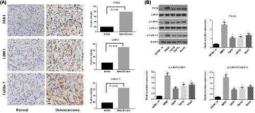
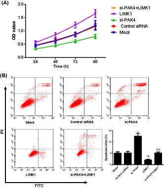
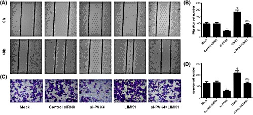
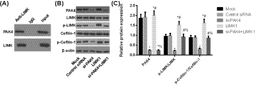
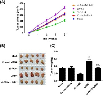
Methods: The expression of PAK4/LIMK1/Cofilin-1 was detected by immunohistochemistry in osteosarcoma tissues. The osteosarcoma cell line MG63 was transfected and divided into Mock, Control siRNA, si-PAK4, LIMK1, and si-PAK4+LIMK1 groups. Then, the cellular biological features of MG63 cells were detected by CCK-8, wound-healing, Transwell, and flow cytometry methods. The relationship of PAK4 and LIMK1 was performed by co-immunoprecipitation test, and the protein expression of PAK4/LIMK1/Cofilin-1 was determined by Western blotting. Finally, the effect of PAK4 on the growth of osteosarcoma was verified by subcutaneous transplantation model of osteosarcoma in nude mice.
Results: The expression of PAK4/LIMK1/Cofilin-1 in both osteosarcoma tissues and cells was up-regulated. Positive PAK4, LIMK1, and Cofilin-1 expressions in osteosarcoma were associated with the clinical stage, distant metastasis, and tumor grade. The MG63 cell viability, migration, and invasion, as well as the expression of PAK4, p-LIMK/LIMK, and p-Cofilin-1/Cofilin-1, were restrained by the knock down of PAK4 while it promoted apoptosis. PAK4 silencing also suppressed the growth of subcutaneous transplanted tumor in nude mice. Co-immunocoprecipitation showed that LIMK and PAK4 protein can form complex in osteosarcoma cells. Besides, LIMK1 overexpression reversed the inhibition effect of PAK4 siRNA on the growth of osteosarcoma cells.
Conclusion: The expression of PAK4/LIMK1/Cofilin-1 pathway in osteosarcoma tissues was up-regulated. Thus, PAK4 inhibition may restrict the osteosarcoma cell proliferation, invasion, and migration but promote its apoptosis via decreasing the activity of LIMK1/Cofilin-1 pathway.
Keywords: Cofilin-1; LIMK1; PAK4; osteosarcoma.
© 2020 The Authors. Journal of Clinical Laboratory Analysis published by Wiley Periodicals LLC.
Conflict of interest statement
The authors declare that there is no conflict of interests regarding the publication of this paper.
Figures
Similar articles
-
DADS downregulates the Rac1-ROCK1/PAK1-LIMK1-ADF/cofilin signaling pathway, inhibiting cell migration and invasion.Oncol Rep. 2013 Feb;29(2):605-12. doi: 10.3892/or.2012.2168. Epub 2012 Dec 6. Oncol Rep. 2013. PMID: 23233092
-
LIMK1 promotes the development of cervical cancer by up-regulating the ROS/Src-FAK/cofilin signaling pathway.Aging (Albany NY). 2024 Jul 5;16(13):11090-11102. doi: 10.18632/aging.206007. Epub 2024 Jul 5. Aging (Albany NY). 2024. PMID: 38975937 Free PMC article.
-
MiR-145 inhibits human colorectal cancer cell migration and invasion via PAK4-dependent pathway.Cancer Med. 2017 Jun;6(6):1331-1340. doi: 10.1002/cam4.1029. Epub 2017 Apr 24. Cancer Med. 2017. PMID: 28440035 Free PMC article.
-
The Role of LIM Kinase in the Male Urogenital System.Cells. 2021 Dec 28;11(1):78. doi: 10.3390/cells11010078. Cells. 2021. PMID: 35011645 Free PMC article. Review.
-
Role of LIMK1-cofilin-actin axis in dendritic spine dynamics in Alzheimer's disease.Cell Death Dis. 2025 Jun 3;16(1):431. doi: 10.1038/s41419-025-07741-7. Cell Death Dis. 2025. PMID: 40461464 Free PMC article. Review.
Cited by
-
Tetramethylpyrazine ameliorates acute lung injury by regulating the Rac1/LIMK1 signaling pathway.Front Pharmacol. 2023 Jan 6;13:1005014. doi: 10.3389/fphar.2022.1005014. eCollection 2022. Front Pharmacol. 2023. PMID: 36686718 Free PMC article.
-
LIM Kinases, LIMK1 and LIMK2, Are Crucial Node Actors of the Cell Fate: Molecular to Pathological Features.Cells. 2023 Mar 4;12(5):805. doi: 10.3390/cells12050805. Cells. 2023. PMID: 36899941 Free PMC article. Review.
-
The role of p21-activated kinase 4 in the progression of oral squamous cell carcinoma by targeting PI3K-AKT signaling pathway.Clin Transl Oncol. 2023 Mar;25(3):739-747. doi: 10.1007/s12094-022-02980-y. Epub 2023 Jan 2. Clin Transl Oncol. 2023. PMID: 36593383
-
Interfering with pak4 Protein Expression Affects Osteosarcoma Cell Proliferation and Migration.Biomed Res Int. 2021 Dec 30;2021:9977001. doi: 10.1155/2021/9977001. eCollection 2021. Biomed Res Int. 2021. Retraction in: Biomed Res Int. 2024 Mar 20;2024:9849782. doi: 10.1155/2024/9849782. PMID: 35005025 Free PMC article. Retracted.
-
LIM Kinases in Osteosarcoma Development.Cells. 2021 Dec 15;10(12):3542. doi: 10.3390/cells10123542. Cells. 2021. PMID: 34944050 Free PMC article. Review.
References
-
- Wu PK, Chen WM, Lee OK, et al. The prognosis for patients with osteosarcoma who have received prior manipulative therapy. J Bone Joint Surg Br. 2010;92(11):1580‐1585. - PubMed
-
- Anderson ME. Update on survival in osteosarcoma. Orthop Clin North Am. 2016;47(1):283‐292. - PubMed
-
- Clark JC, Dass CR, Choong PF. A review of clinical and molecular prognostic factors in osteosarcoma. J Cancer Res Clin Oncol. 2008;134(3):281‐297. - PubMed
-
- Janeway KA, Grier HE. Sequelae of osteosarcoma medical therapy: a review of rare acute toxicities and late effects. Lancet Oncol. 2010;11(7):670‐678. - PubMed
-
- Kushlinskii NE, Fridman MV, Braga EA. Molecular mechanisms and microRNAs in osteosarcoma pathogenesis. Biochemistry (Moscow). 2016;81(4):315‐328. - PubMed
MeSH terms
Substances
LinkOut - more resources
Full Text Sources
Medical
Research Materials
