

Does contrast-enhanced ultrasound (CEUS) play a better role in diagnosis of breast lesions with calcification? A comparison with MRI
- PMID: 32463295
- PMCID: PMC7446019
- DOI: 10.1259/bjr.20200195
Does contrast-enhanced ultrasound (CEUS) play a better role in diagnosis of breast lesions with calcification? A comparison with MRI
Abstract
Objective: To compare the efficacy of contrast-enhanced ultrasound enabled reclassification of Breast Imaging Reporting and Data System (CEUS-BI-RADS) with MRI in the diagnosis of breast lesions with calcification.
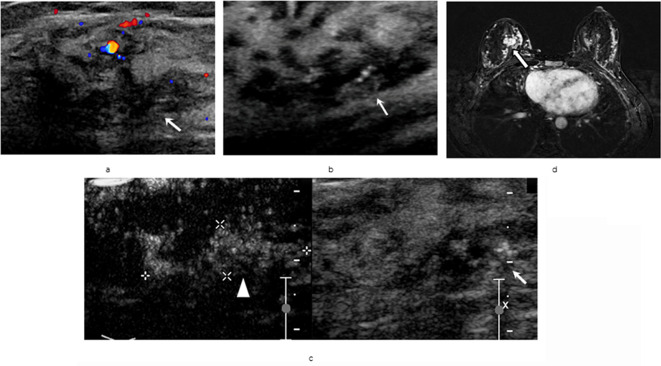
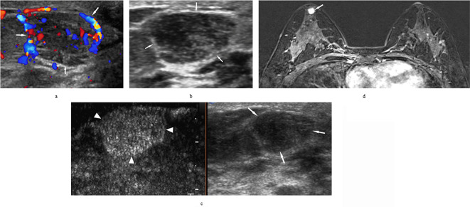
Methods: A total of 52 breast lesions with calcification from 51 patients were detected by ultrasound as hyperechoic foci and categorized as BI-RADS 3-5. The 51 patients further underwent CEUS scan and MRI. The ultrasound-BI-RADS combined with CEUS 5-point score system redefined the classification of BI-RADS which was called CEUS-BI-RADS. The diagnostic efficacy of three methods was assessed by receiver operating characteristic (ROC) curve analysis. Histopathological assessment used as the gold-standard.
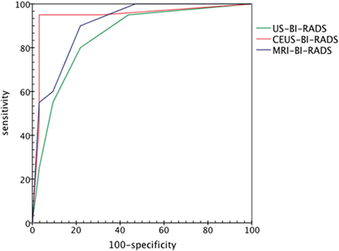
Results: The sensitivities of Ultrasound-BI-RADS, MRI classification of BI-RADS (MRI-BI-RADS) and CEUS-BI-RADS were 85%, 90% and 95% without significant difference among the three modalities (p > 0.05). The diagnostic specificities of ultrasound-BI-RADS, MRI-BI-RADS and CEUS-BI-RADS were 78.1%, 78.1% and 96.8%, respectively (p < 0.05); and the accuracy were 80.7%, 82.6% and 96.1% for ultrasound-BI-RADS, MRI-BI-RADS and CEUS-BI-RADS, respectively (p < 0.05). The area under ROC (AUROC) in differentiation of breast lesions with calcification was 0.945 for CEUS-BI-RADS, 0.907 for MRI-BI-RADS and 0.853 for ultrasound-BI-RADS, with no significant difference among the three modalities (p > 0.05).
Conclusion: The CEUS-BI-RADS has a better diagnostic efficiency than MRI-BI-RADS in the differentiation of the breast lesions with calcification.
Advances in knowledge: •CEUS is a better method in differentiation of breast lesions with calcification.•CEUS-BI-RADS increases the efficiency of diagnosis compared to MRI.
Figures
References
-
- Bonfiglio R, Scimeca M, Toschi N, Pistolese CA, Giannini E, Antonacci C, et al. Radiological, histological and chemical analysis of breast microcalcifications: diagnostic value and biological significance. J Mammary Gland Biol Neoplasia 2018; 23(1-2): 89–99. doi: 10.1007/s10911-018-9396-0 - DOI - PubMed
-
- Du J, Wang L, Wan C-F, Hua J, Fang H, Chen J, et al. Differentiating benign from malignant solid breast lesions: combined utility of conventional ultrasound and contrast-enhanced ultrasound in comparison with magnetic resonance imaging. Eur J Radiol 2012; 81: 3890–9. doi: 10.1016/j.ejrad.2012.09.004 - DOI - PubMed
Publication types
MeSH terms
Substances
LinkOut - more resources
Full Text Sources
Medical
