. 2020 Jul 1;319(1):G11-G22.
doi: 10.1152/ajpgi.00098.2020. Epub 2020 May 28.

Lipid droplets disrupt mechanosensing in human hepatocytes

Affiliations

Lipid droplets disrupt mechanosensing in human hepatocytes

LiKang Chin et al. Am J Physiol Gastrointest Liver Physiol. .

Abstract

Hepatocellular carcinoma (HCC) is the fourth-leading cause of cancer death in the world. Although most cases occur in stiff, cirrhotic livers, and stiffness is a significant risk factor, HCC can also arise in noncirrhotic livers in the setting of nonalcoholic fatty liver disease (NAFLD). We hypothesized that lipid droplets in NAFLD might apply mechanical forces to the nucleus, functioning as mechanical stressors akin to stiffness. We investigated the effect of lipid droplets on cellular mechanosensing and found that primary human hepatocytes loaded with the fatty acids oleate and linoleate exhibited decreased stiffness-induced cell spreading and disrupted focal adhesions and stress fibers. The presence of large lipid droplets in hepatocytes resulted in increased nuclear localization of the mechano-sensor Yes-associated protein (YAP). In cirrhotic livers from patients with NAFLD, hepatocytes filled with large lipid droplets showed significantly higher nuclear localization of YAP as compared with cells with small lipid droplets. This work suggests that lipid droplets induce a mechanical signal that disrupts the ability of the hepatocyte to sense its underlying matrix stiffness and that the presence of lipid droplets can induce intracellular mechanical stresses.NEW & NOTEWORTHY This work examines the impact of lipid loading on mechanosensing by human hepatocytes. In cirrhotic livers, the presence of large (although not small) lipid droplets increased nuclear localization of the mechanotransducer YAP. In primary hepatocytes in culture, lipid droplets led to decreased stiffness-induced cell spreading and disrupted focal adhesions and stress fibers; the presence of large lipid droplets resulted in increased YAP nuclear localization. Collectively, the data suggest that lipid droplets induce intracellular mechanical stress.

Keywords: HCC; NAFLD; YAP; cirrhosis; lipid droplet.


Conflict of interest statement

No conflicts of interest, financial or otherwise, are declared by the authors.

Figures

None
Graphical abstract
Fig. 1.
Fig. 1.
Nonalcoholic fatty liver disease (NAFLD) livers have more lipid but similar stiffness and collagen as non-NAFLD cirrhotic livers. A: representative images of hematoxylin and eosin (H & E)-, Sirius Red-, and Oil Red O (ORO)-stained human livers staged as intermediate/advanced cirrhosis from patients with or without a documented clinical history of NAFLD. Livers exhibited nodules surrounded by fibrous septa(*); those with NAFLD also had large lipid droplets (arrows). B: shear storage modulus G′ of human livers from patients with or without a history of NAFLD. C: collagen content, as determined by %Sirius Red staining, in cirrhotic livers was similar with or without NAFLD. D: NAFLD livers had more ORO staining than non-NAFLD livers (P = 0.027). E and F: scatter plots of shear storage modulus G′ vs. %Sirius Red staining showing nonlinear correlation (power law fit, P = 0.022; E) and %ORO staining showing no correlation (power law fit, P = 0.98; F). Scale bar, 50 µm. Error bars are SE. Shear storage modulus G′ and Sirius Red: n = 27 non-NAFLD and 9 NAFLD. ORO: n = 5 non-NAFLD and 6 NAFLD.
Fig. 2.
Fig. 2.
Nuclear Yes-associated protein (YAP) localization is increased in hepatocytes with large lipid droplets. A: representative images of human livers with intermediate or advanced cirrhosis, with or without nonalcoholic fatty liver disease (NAFLD), stained for YAP. Individual cells from 5 livers were categorized as having small or large lipid droplets. Hepatocytes with no lipid droplets or with small lipid droplets exhibited round nuclei that were both YAP+ (black arrows) and YAP− (white arrows). Hepatocytes with large lipid droplets exhibited deformed nuclei that were almost exclusively YAP+ (black arrows). B: quantification showed that hepatocytes with large lipid droplets had significantly more nuclear YAP than cells with small droplets or lipid droplet-free cells in nonsteatotic cirrhotic livers. Scale bar, 50 µm. **P ≤ 0.01; ***P ≤ 0.001. Error bars are SE. Each data point represents cells from 1 liver; n = 5 control (cirrhotic) livers (116–340 cells/data point) and 5 NAFLD livers (4 of the 5 had hepatocytes with small droplets, 13–104 cells counted/data point, whereas all 5 had hepatocytes with large droplets, 30–164 cells counted/data point). One NAFLD liver had <10 cells with small droplets in the fields examined and was dominated by large droplets; it was excluded from the NAFLD, small-droplet group.
Fig. 3.
Fig. 3.
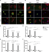
Oleate-treated cells store more lipid than cells treated with linoleate, and lipid storage decreases with stiffness. Representative confocal images of primary human hepatocytes (PHH; A) and HuH7 cells (B) seeded on 500-Pa (soft) or 10-kPa (stiff) polyacrylamide (PAA)-collagen gels or glass (collagen-coated for PHH), treated with 400 µM oleate or linoleate for 48 h, and stained for neutral lipids (green), actin (red), and nuclei (blue). BODIPY mean intensity (a measure of lipid density; C and D) and integrated density (a measure of total lipid; E and F) were quantified for PHH and HuH7. Oleate-treated HuH7 cells had significantly less lipid density on soft gels and glass (P ≤ 0.01, not graphically represented; C and D) and less total lipid on all substrates (P ≤ 0.0001, not graphically represented; E and F) than their normal PHH counterparts. To better visualize lipid droplets in BSA and linoleate (only PHH) groups, gain was increased all to the same setting; gain was increased in BSA-treated PHH to better see actin. All quantification was performed on images acquired with the same settings. Scale bar, 50 µm. *P ≤ 0.05, **P ≤ 0.01, ***P ≤ 0.001, and ****P ≤ 0.0001. PHH, n = 30 total cells/condition from three independent experiments using cells from 2 donors. HuH7, n = 12–25 total cells/condition from 2 independent experiments. Error bars are ± SE.
Fig. 4.
Fig. 4.
Stiffness-dependent cell spreading decreases in fatty acid-loaded primary human hepatocytes (PHH). A: representative live-cell bright-field images of PHH seeded on 500-Pa (soft) and 10-kPa (stiff) PAA-collagen gels or collagen-coated glass after 48 h of treatment with 400 µM oleate or linoleate. BG: cell area (B and D), circularity (C and E), and solidity (F and G) were quantified for both PHH and HuH7 cells (images shown in Supplemental Fig. S3). Scale bar, 50 µm. *P ≤ 0.05; **P ≤ 0.01; ***P ≤ 0.001; ****P ≤ 0.0001; #0.05<P ≤ 0.10 (n = 60 total cells/condition from 3 independent experiments using cells from one donor). Error bars are ± SE.
Fig. 5.
Fig. 5.
Fatty acid treatment disrupts stress fibers and focal adhesions in primary human hepatocytes (PHH), particularly on glass. A: representative confocal images of PHH seeded on 500-Pa (soft) or 10-kPa (stiff) polyacrylamide (PAA)-collagen gels, or collagen-coated glass, treated with 400 µM oleate or linoleate for 48 h and stained for vinculin (green), actin (red), and nuclei (blue). BD: quantification is shown for %cells that exhibited stress fibers (B), vinculin patches, as defined by punctate staining located at the end of stress fibers (C), and total cell area of vinculin staining (except for oleate-treated cells due to nonspecific staining of the lipid droplets; D). Intensity was optimized for each image to best visualize the vinculin and actin staining (B and C). Quantification of vinculin staining (D) was from the original images, all taken at the same settings, as shown in Supplemental Fig. S4A. Scale bar, 50 µm. *P ≤ 0.05; **P ≤ 0.01; ****P ≤ 0.0001; #0.05<P ≤ 0.100. %Stress fibers or vinculin, n = 3 independent experiments. Vinculin area, n = 32–63 total cells/condition from 3 independent experiments using cells from 1 donor. Error bars are ± SE.
Fig. 6.
Fig. 6.
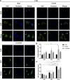
Cells with small lipid droplets show no change in Yes-associated protein (YAP) nuclear translocation or mean intensity. A: representative confocal images of primary human hepatocytes (PHH) seeded on 500-Pa (soft) or 10-kPa (stiff) PAA-collagen gels, or collagen-coated glass, treated with 400 µM oleate or linoleate for 48 h, and stained for YAP (green) and nuclei (blue). B: nuclear-to-cytosolic YAP ratio was quantified. C: YAP mean intensity of the entire cell was measured. Although YAP intensity exhibited some stiffness dependence, fatty acid treatment had no effect. Scale bar, 50 µm. *P ≤ 0.05; **P ≤ 0.01; ***P ≤ 0.001; ****P ≤ 0.0001; #0.05<P ≤ 0.100; n = 29–53 total cells/condition from 3 independent experiments using cells from 2 donors. Error bars are ± SE.
Fig. 7.
Fig. 7.
Large lipid droplets are associated with increased nuclear Yes-associated protein (YAP) in cells on glass. A: representative confocal images of primary human hepatocytes (PHH) seeded on 500-Pa (soft) or 10-kPa (stiff) polyacrylamide (PAA)-collagen gels or collagen-coated glass treated with either 400 µM oleate to generate small lipid droplets or a combination of 400 µM oleate and 100 nM insulin for large lipid droplets. Cells were stained for lipid (green), YAP (red), and nuclei (blue). Intensity was optimized for each stiffness to best visualize YAP staining. Nuclei alone are shown in insets. Cells from both treatment conditions were binned into those with predominantly small, medium, or large lipid droplets. B and C: the nuclear-to-cytosolic YAP ratio (B) and nuclear circularity (C) were quantified for cells with small vs. large droplets. Cells with large lipid droplets had significantly greater nuclear YAP and greater nuclear deformation than those with small lipid droplets when seeded on glass. Inset scale bar, 10 µm. Scale bar, 50 µm. *P ≤ 0.05; **P ≤ 0.01; ***P ≤ 0.001; ****P ≤ 0.0001 (n = 22–39 total cells/condition from 4 independent experiments using cells from 1 donor). Error bars are ± SE.

Similar articles

Cited by

References

    1. Adler M, Larocca L, Trovato FM, Marcinkowski H, Pasha Y, Taylor-Robinson SD. Evaluating the risk of hepatocellular carcinoma in patients with prominently elevated liver stiffness measurements by FibroScan: a multicentre study. HPB (Oxford) 18: 678–683, 2016. doi:10.1016/j.hpb.2016.05.005. - DOI - PMC - PubMed
    1. Baker A-M, Bird D, Lang G, Cox TR, Erler JT. Lysyl oxidase enzymatic function increases stiffness to drive colorectal cancer progression through FAK. Oncogene 32: 1863–1868, 2013. doi:10.1038/onc.2012.202. - DOI - PubMed
    1. Baker BM, Trappmann B, Wang WY, Sakar MS, Kim IL, Shenoy VB, Burdick JA, Chen CS. Cell-mediated fibre recruitment drives extracellular matrix mechanosensing in engineered fibrillar microenvironments. Nat Mater 14: 1262–1268, 2015. doi:10.1038/nmat4444. - DOI - PMC - PubMed
    1. Boyd NF, Li Q, Melnichouk O, Huszti E, Martin LJ, Gunasekara A, Mawdsley G, Yaffe MJ, Minkin S. Evidence that breast tissue stiffness is associated with risk of breast cancer. PLoS One 9: e100937, 2014. doi:10.1371/journal.pone.0100937. - DOI - PMC - PubMed
    1. Caliari SR, Perepelyuk M, Cosgrove BD, Tsai SJ, Lee GY, Mauck RL, Wells RG, Burdick JA. Stiffening hydrogels for investigating the dynamics of hepatic stellate cell mechanotransduction during myofibroblast activation. Sci Rep 6: 21387, 2016. doi:10.1038/srep21387. - DOI - PMC - PubMed

Publication types

MeSH terms