. 2020 Jul 9;48(12):6624-6639.
doi: 10.1093/nar/gkaa406.

MEIOK21: a new component of meiotic recombination bridges required for spermatogenesis

Affiliations

MEIOK21: a new component of meiotic recombination bridges required for spermatogenesis

Yongliang Shang et al. Nucleic Acids Res. .

Abstract

Repair of DNA double-strand breaks (DSBs) with homologous chromosomes is a hallmark of meiosis that is mediated by recombination 'bridges' between homolog axes. This process requires cooperation of DMC1 and RAD51 to promote homology search and strand exchange. The mechanism(s) regulating DMC1/RAD51-ssDNA nucleoprotein filament and the components of 'bridges' remain to be investigated. Here we show that MEIOK21 is a newly identified component of meiotic recombination bridges and is required for efficient formation of DMC1/RAD51 foci. MEIOK21 dynamically localizes on chromosomes from on-axis foci to 'hanging foci', then to 'bridges', and finally to 'fused foci' between homolog axes. Its chromosome localization depends on DSBs. Knockout of Meiok21 decreases the numbers of HSF2BP and DMC1/RAD51 foci, disrupting DSB repair, synapsis and crossover recombination and finally causing male infertility. Therefore, MEIOK21 is a novel recombination factor and probably mediates DMC1/RAD51 recruitment to ssDNA or their stability on chromosomes through physical interaction with HSF2BP.


Figures

Figure 1.
Figure 1.
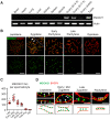
Dynamic localization of MEIOK21 on meiotic chromosomes. (A) Meiok21 is highly expressed in testes and fetal ovaries, revealed by RT-qPCR. (B) The dynamic localization of MEIOK21 to meiotic chromosomes in mouse spermatocytes. The meiotic stages of spermatocytes were determined by the SYCP3 staining (red) of the chromosome axis. MEIOK21 (green) are visualized on meiotic chromosomes from leptotene to late pachytene and are undetectable in diplotene. The arrow in zygotene indicates MEIOK21 foci on an unsynapsed axis, 77.60 ± 8.24% in early/mid zygotene and 26.08 ± 8.98% in late zygotene. The arrowhead indicates MEIOK21 foci on a synapsed axis, 22.40 ± 8.24% in early/mid zygotene and 73.92 ± 8.98% in late zygotene. Mean ± SD, n = 20 for each group. The dashed line separates the target cell from the adjacent unrelated cell. Scale bar, 10 μm. (C) Quantification of the numbers of MEIOK21 foci in spermatocytes from leptotene to diplotene. Spermatocyte sample sizes, n = 20 for each stage. Lep, leptotene; early Zyg, early zygotene; late Zyg, late zygotene; early Pac, early pachytene; late Pac, late pachytene; Dip, diplotene. Error bar, mean ± SD. (D) The dynamic localization of MEIOK21 on chromosomes investigated by structured illumination microscope (SIM). (Top panel) MEIOK21 foci first localize on the chromosome axis (‘leptotene’, arrows). Then, on chromosome regions where two homolog axes are aligned in parallel, MEIOK21 foci were frequently seen as ‘pairs’ at the opposing sites on the two homologs (‘early/mid zygotene’, arrows, MEIOK21 foci; blue dotted lines, focus pairs) and some foci are released as ‘hanging foci’ (‘early/mid zygotene’, arrowheads). When homologous chromosomes aligned closely, MEIOK21 appears as ‘bridges’ or ‘fusing foci’ (‘late zygotene’, arrowhead) and finally as fused foci (‘pachytene’, arrowheads) between synapsed axes. (Bottom panel) cartoons illustrate the dynamic localization of MEIOK21 with meiotic progression. Scale bar, 1 μm.
Figure 2.
Figure 2.
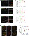
MEIOK21 localizes to meiotic recombination sites in a DSB dependent manner. (A) MEIOK21 colocalizes with RPA. (B) Quantification shows that ∼85-95% of the two factors overlap with each other from leptotene to early pachytene. Error bar, mean ± SD. Sample sizes (from left to right), n = 13, 13, 37, 37, 30 and 30, respectively. (C) MEIOK21 partially colocalizes with DMC1. (D) From leptotene to zygotene, ∼40% of the two factors overlap with each other; in early pachytene, ∼20% of MEIOK21 foci overlap with DMC1 foci, and ∼30% of DMC1 foci overlap with MEIOK21 foci. Error bar, mean ± SD. Sample sizes (from left to right), n = 30, 30, 41, 41, 30 and 30, respectively. Arrowheads indicate overlapped foci. (E) MEIOK21 partially colocalizes with RAD51. (F) From leptotene to zygotene, ∼60% of the two factors overlap with each other; in early pachytene, ∼20% of MEIOK21 foci overlap with RAD51 foci, and ∼30% of RAD51 foci overlap with MEIOK21 foci. Error bar, mean ± SD. Sample sizes (from left to right), n = 17, 17, 42, 42, 30 and 30, respectively. Arrowheads indicate overlapped foci. (G) MEIOK21 foci are abolished in Spo11/ spermatocytes, but more MEIOK21 foci are observed in Dmc1/ spermatocytes compared to WT. Note that Spo11/ and Dmc1/ spermatocytes can only reach the ‘zygotene’ stage. The dashed line separates the target cell from the adjacent unrelated cell. Scale bar, 10 μm (A, C, E, G). (H) The quantification of MEIOK21 foci in spermatocytes of WT, Spo11/ and Dmc1/ mice. Spermatocyte sample sizes, from left to right, n = 19, 22, 17, 20, 22 and 21. Error bar, mean ± SD.
Figure 3.
Figure 3.
MEIOK21 interacts with recombination factor HSF2BP. (A) MEIOK21 interacts with HSF2BP in Y2H screening, with MEIOK21 as prey and HSF2BP as bait. About 105 yeast cells were seeded in each dot. The experiment was repeated three times. (B) HSF2BP is pulled-down in Meiok21+/+ testis lysate (+/+) (200 mg) but not Meiok21/ testis lysate (−/−) (200 mg) by antibody against MEIOK21.The input amount is ∼5% of the lysate. IgG acts as a negative control. The experiment was repeated three times. Nonspecific bands are labeled with *. (C) MEIOK21 colocalizes with HSF2BP on meiotic chromosomes (see Supplementary Figure 2E for quantifications). Scale bar, 10 μm. (D) Diagram of MEIOK21 truncations. (E) The C terminus of MEIOK21 (a.a. 451–600) is required for interaction with HSF2BP. FLAG-tagged HSF2BP and GFP-tagged MEIOK21 truncations were co-transfected to HEK-293T cells. After 48 hours, cells were lysed, and co-IP experiments were performed using GFP antibody. The input amount is ∼5% of the lysate. The experiment was repeated twice. (F) The N terminus of MEIOK21 (a.a. 1–150) is required for its nuclear localization. GFP-tagged MEIOK21 truncations were transfected to HEK-293T cells. After 24 hours, GFP signal was examined under a fluorescence microscope. Scale bar, 5 μm. (G) The diagram of HSF2BP truncations. (H) The coiled-coil domain of HSF2BP (a.a. 1–92) is required for MEIOK21 binding. GFP-tagged MEIOK21 and FLAG-tagged HSF2BP truncations were co-transfected to HEK-293T cells. After 48 hours, cells were lysed, and co-IP experiments were performed with antibodies against GFP. The input amount is ∼5% of the lysate. The experiment was repeated twice. The non-specific band (*). (I) Diagrams to illustrate interaction domains of MEIOK21 and HSF2BP.
Figure 4.
Figure 4.
MEIOK21 is required for male fertility. (A) The schematic of Meiok21 knockout allele. Exons 3–6 were deleted in the mutant. (B) Meiok21 knockout was confirmed by RT-PCR (Materials and Methods). RT-PCR was performed using cDNA obtained from testes as templates. PCR products with proper size were detected from WT but not mutant samples. (C) Meiok21/ male mice were sterile. n = 5 for each genotype. Error bar, mean ± SEM. (DE) Both the testis size and testis/body weight ratio of 8-week-old Meiok21/ mice are significantly reduced compared to Meiok21+/+and Meiok21+/ mice. Error bar, mean ± SEM. n = 3 for each genotype. (F) No spermatids or mature spermatozoa were observed in testis or epididymis by HE staining in Meiok21/ mice. Scale bar, 50 μm. (G) A large number of apoptotic spermatocytes (green) were observed in Meiok21/ testis by TUNEL staining. Scale bar, 50 μm. (H) Quantitative analysis of spermatocytes at different meiotic stages from Meiok21+/+ and Meiok21/ mice testes at postnatal day 20. There are more zygotene but less pachytene spermatocytes in Meiok21/ testis compared to WT. n = 3 for each genotype. Error bar, mean ± SEM.
Figure 5.
Figure 5.
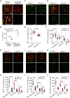
MEIOK21 is required for efficient meiotic recombination. (A) Two types of aberrant synapsis are frequently observed in Meiok21/ spermatocytes, as seen in other recombination defective mutants. Partner switch (left panel), i.e. one chromosome (arrow) synapses with more than one partner (arrowheads). (B) Quantification of the two types of aberrant ‘zygotene’ nuclei described in (A). For each genotype, 3 mice were examined. The numbers of cells examined in each mouse are 84, 60 and 60 for WT, and 93, 48 and 64 for Meiok21/. (CD) The number of crossovers, marked by MLH1 foci (red), is significantly reduced in Meiok21/ pachytene spermatocytes compared to WT. n = 20 and 23 for WT and Meiok21/, respectively. Chromosomes without even one MLH1 focus (arrows). (E, F) In Meiok21/ spermatocytes, the level of RPA foci is similar to that of WT at zygotene, but there is aberrant retention of RPA on chromosomes at a late time. From left to right, n = 20, 20, 24, 13, 23, 19, 14 and 20, respectively. (G–L) The numbers of DMC1, RAD51 and HSF2BP foci are greatly decreased in Meiok21/ spermatocytes compared to WT. Early/Mid Zyg, early or middle zygotene; Late Zyg, late zygotene; Pac, pachytene, Late Pac: late pachytene, Dip: diplotene. For each panel from left to right, n = 26, 36, 24, 30, 22 and 27 (H), 29, 36, 20, 27, 29 and 31 (J), 20, 21, 22, 21, 21 and 21 (L). Scale bar, 10 μm (A, C, E, G, I, K). Error bar, mean ± SD (D, F, H, J, L) or mean ± SEM (B).

Similar articles

Cited by

References

    1. Nagaoka S.I., Hassold T.J., Hunt P.A.. Human aneuploidy: mechanisms and new insights into an age-old problem. Nat. Rev. Genet. 2012; 13:493–504. - PMC - PubMed
    1. Capalbo A., Hoffmann E.R., Cimadomo D., Ubaldi F.M., Rienzi L.. Human female meiosis revised: new insights into the mechanisms of chromosome segregation and aneuploidies from advanced genomics and time-lapse imaging. Hum. Reprod. Update. 2017; 23:706–722. - PubMed
    1. Wang S., Liu Y., Shang Y., Zhai B., Yang X., Kleckner N., Zhang L.. Crossover interference, crossover maturation, and human aneuploidy. Bioessays. 2019; 41:e1800221. - PMC - PubMed
    1. Zickler D., Kleckner N.. Recombination, pairing, and synapsis of homologs during meiosis. Cold Spring Harb. Perspect. Biol. 2015; 7:a016626. - PMC - PubMed
    1. Keeney S., Giroux C.N., Kleckner N.. Meiosis-specific DNA double-strand breaks are catalyzed by Spo11, a member of a widely conserved protein family. Cell. 1997; 88:375–384. - PubMed

Publication types

MeSH terms