. 2020 May 28;16(5):e1007926.
doi: 10.1371/journal.pcbi.1007926. eCollection 2020 May.

In silico analysis of hypoxia activated prodrugs in combination with anti angiogenic therapy through nanocell delivery

Affiliations

In silico analysis of hypoxia activated prodrugs in combination with anti angiogenic therapy through nanocell delivery

Cameron Meaney et al. PLoS Comput Biol. .

Abstract

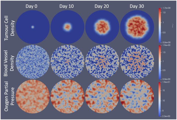
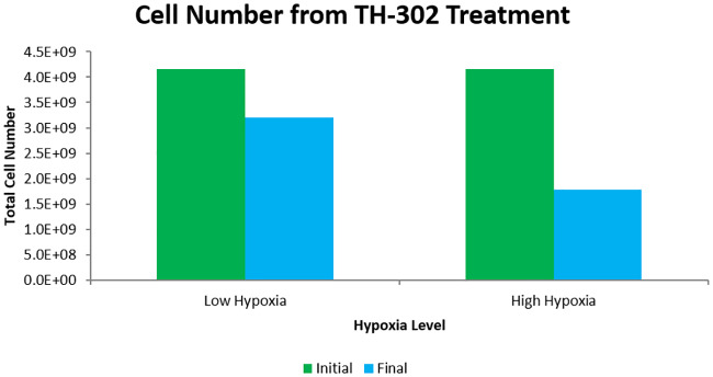
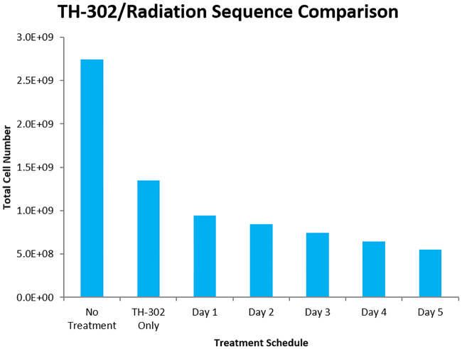
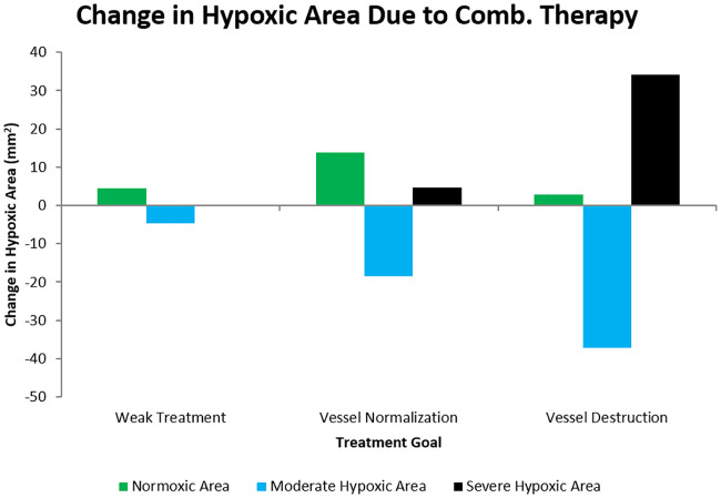
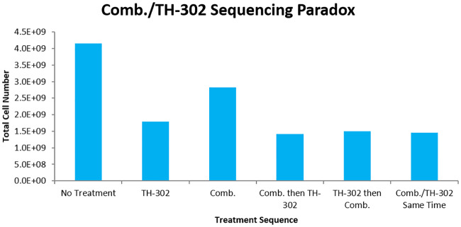
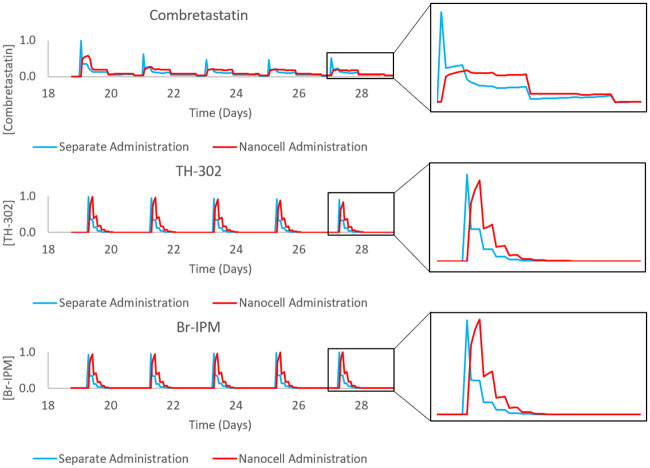
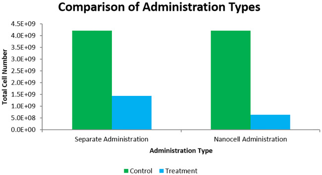
Tumour hypoxia is a well-studied phenomenon with implications in cancer progression, treatment resistance, and patient survival. While a clear adverse prognosticator, hypoxia is also a theoretically ideal target for guided drug delivery. This idea has lead to the development of hypoxia-activated prodrugs (HAPs): a class of chemotherapeutics which remain inactive in the body until metabolized within hypoxic regions. In theory, these drugs have the potential for increased tumour selectivity and have therefore been the focus of numerous preclinical studies. Unfortunately, HAPs have had mixed results in clinical trials, necessitating further study in order to harness their therapeutic potential. One possible avenue for the improvement of HAPs is through the selective application of anti angiogenic agents (AAs) to improve drug delivery. Such techniques have been used in combination with other conventional chemotherapeutics to great effect in many studies. A further benefit is theoretically achieved through nanocell administration of the combination, though this idea has not been the subject of any experimental or mathematical studies to date. In the following, a mathematical model is outlined and used to compare the predicted efficacies of separate vs. nanocell administration for AAs and HAPs in tumours. The model is experimentally motivated, both in mathematical form and parameter values. Preliminary results of the model are highlighted throughout which qualitatively agree with existing experimental evidence. The novel prediction of our model is an improvement in the efficacy of AA/HAP combination therapies when administered through nanocells as opposed to separately. While this study specifically models treatment on glioblastoma, similar analyses could be performed for other vascularized tumours, making the results potentially applicable to a range of tumour types.


Conflict of interest statement

The authors have declared that no competing interests exist.

Figures

Fig 1
Fig 1. Cases of tumour vascularization.
In A. the tumour is overvascularized with chaotic, irregular vessels (m > 1) causing perfusion-limited hypoxia. In B. vascular normalization (m ≈ 1) is achieved where optimal oxygen or drug extravasation is realized. In C. a strong dose of AAs causes vascular destruction (m < 1), leading to diffusion-limited hypoxia in the tumour. The quantity m is the tumour blood vessel density.
Fig 2
Fig 2. Delivery rate from vessels as a function of the average vessel density.
Curve is described by the function me(mmlim)2. Notice that the optimal vessel delivery occurs at m = 1. As m becomes greater than 1, the delivery rate decreases, allowing for the impaired delivery due to overvascularization.
Fig 3
Fig 3. Visualization of model results for tumour cell density, blood vessel density, and oxygen partial pressure over a 30 day span with no treatment.
Model is solved using the FEniCS Project Finite Element Solver [55, 56] and is visualized in Paraview [57]. Tumour cells begin as a normalized Gaussian distribution with a standard deviation of approximately 0.63mm. Vessels start randomly distributed between 0 and 1 over the domain. Oxygen is solved in quasi-steady state with respect to the time step length. Day 0 corresponds to the solution of the system after the first time step. Notice that vessels grow toward islands or tubes of density 0 and 1 in the absence of tumour cells, but will become overvascularized (m > 1) in the presence of tumour cells. Also notice that the oxygen concentration decreases in tumour area due to the overvascularization. Scales on the right correspond to the nondimensional units.
Fig 4
Fig 4. Comparison of TH-302 effectiveness for tumours of different hypoxic levels.
The tumour is allowed to grow for 15 days before TH-302 treatment. TH-302 is given every other day starting on the 15th day and the plasma concentration is assumed to have a half life of 3 hours. The high hypoxic case is from the parameters listed in Table 1 while the low hypoxia case assumes a halved oxygen supply rate (rk). Initial cell number is taken from the time step immediately before treatment and final cell number is taken from the time step immediately following treatment. TH-302 treatment kills only 23% of tumour cells in the low hypoxic case while it kills 57% of cells in the high hypoxic case.
Fig 5
Fig 5. Efficacy of TH-302/radiotherapy combinations based on administration sequence.
The tumour is allowed to grow for 15 days with TH-302 treatment starting on the 15th day. TH-302 is then given every day for 5 straight days with radiation given 6 hours after TH-302 on one of the days. Resulting cell number is calculated at the end of the 5th day, after all treatment has ceased. Notice that the later radiotherapy is administered, the more tumour cells are killed.
Fig 6
Fig 6. Cases of anti angiogenic therapy effect based on combretastatin strength.
Weak treatment corresponds to an anti angiogenic strength parameter of σ = 20, vessel normalization to σ = 30, and vessel destruction to σ = 70. Normoxic area is where [O2] > 10mmHg, moderate hypoxic where 5 < [O2] < 10mmHg, and severe hypoxic where [O2] < 5mmHg. The hypoxic ranges were chosen from the understood ranges of tumour hypoxia ([7, 25] for example). Combretastatin is given every other day starting on the 15th day in decreasing amounts (of magnitudes 1, 0.75, 0.5, 0.25, and 0.1). Notice that there is an optimal middle strength where the increase in normoxic area is maximized.
Fig 7
Fig 7. Combretastatin/TH-302 sequencing paradox with vessel normalization.
The tumour is allowed to grow for 18 days before the start of treatment. Five doses of drugs are given in 2 day increments, each at the same strength. If both types of drugs are given non-simultaneously, then there is a 6 hour delay between their administrations. The optimal schedule is to give combretastatin 6 hours prior to TH-302, although it is only slightly better than other sequences. Furthermore, the combination therapy fails to show any drug synergy effects.
Fig 8
Fig 8. Normalized drug concentrations in the tumour area over the treatment schedule for combretastatin-first treatment administered either separately or through nanocells.
The left column of plots is the full treatment schedule used in simulations, the right column is a zoomed-in picture of the final drug administration to better observe the difference in release profiles. Notice the delayed release in the nanocell administration case.
Fig 9
Fig 9. Comparison of optimal separate administration sequence vs. nanocell administration.
Treatment schedules are as shown in Fig 7. Notice the improvement in cell kill in the nanocell administration.

References

    1. Cosse JP, Michiels C. Tumour Hypoxia Affects the Responsiveness of Cancer Cells to Chemotherapy and Promotes Cancer Progression. Anti-Cancer Agents in Medicinal Chemistry. 2008; 8(7), 790–797. - PubMed
    1. Graeber TG, Osmanian C, Jacks T, Housman DE, Koch CJ, Lowe SW, et al. Hypoxia-mediated selection of cells with diminished apoptotic potential in solid tumours. Nature 379, 88–91 (1996) - PubMed
    1. Graham K. Overcoming tumor hypoxia as a barrier to radiotherapy, chemotherapy and immunotherapy in cancer treatment. International Journal of Nanomedicine. 2018; p. 6049–6058. 10.2147/IJN.S140462 - DOI - PMC - PubMed
    1. Hammond EM, Asselin MC, Forster D, O’Connor JPB, Senra JM, Williams KJ. The Meaning, Measurement and Modification of Hypoxia in the Laboratory and the Clinic. Clinical Oncology. 2014;26(5):277–288. - PubMed
    1. Harris AL. Hypoxia—a Key Regulatory Factor in Tumour Growth. Nature Reviews Cancer. 2002;2(1):38–47. - PubMed

Publication types

Grants and funding