Review
. 2020 May 28;16(5):e1008542.
doi: 10.1371/journal.ppat.1008542. eCollection 2020 May.

Capsid protein is central to the birth of flavivirus particles

Affiliations
Review

Capsid protein is central to the birth of flavivirus particles

Ter Yong Tan et al. PLoS Pathog. .
No abstract available


Conflict of interest statement

The authors have declared that no competing interests exist.

Figures
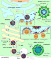
Fig 1
Fig 1. A schematic diagram of the flavivirus maturation pathway.
(A) The viral RNA is translated on ER to produce polypeptide. This polypeptide is digested by host peptidase and viral proteases to form 10 flavivirus proteins, including the structural proteins: E, prM, and C. In the lumen side of ER, E and prM form a heterodimer, and 3 of these heterodimers interact with each other, forming an inverted tripod structure. The tripods then interact with each other. On the cytoplasm side, a C dimer binds to the TM regions of a prM–E tripod and also the viral RNA genome. One C dimer then interacts with 2 other nearby C dimers via their α5 helices to form a triangular network. The end result is one side of the C dimers interacts with lipid bilayer/TM regions of prM–E, while the other interacts with RNA. The flavivirus particle is assembled on the ER membrane, and it then buds off as spiky immature particles. (B) The immature particles are transported through the TGN. A conformational change of the virion from spiky to smooth surface morphology is triggered by the exposure to the more acidic pH environment of the TGN. The virus surface conformational changes expose the furin-cleavage site on the prM, allowing the prM to be cleaved into pr and M proteins. (C) The cleaved pr molecule remains attached to the viral particles at the low pH environment of the exosomes. (D) When released into the extracellular environment, the increased in pH causes the release of pr molecules from the virus surface, forming the fully infectious virus particle. (E) The central slice of the cryoEM density map of immature particles, showing the C dimers bridging the inner leaflet of the lipid bilayer and the RNA genome. (F) The center slice of the cryoEM density map of the mature virus particles, showing a lack of C dimer density at the interface between the inner leaflet of the bilipid layer membrane and the RNA genome. We speculate that the dramatic rearrangement of the E and prM/M proteins during the virus maturation process may result in the release of the C dimers from the inner leaflet of lipid bilayer membrane. The approximate luminal pH values of the cellular compartments are shown [28]. C, Capsid; cryoEM, cryo-electron microscopy; E, Envelope; ER, endoplasmic reticulum; prM, precursor Membrane; TGN, trans-Golgi network; TM, transmembrane.
Fig 2
Fig 2. The organization of structural proteins in flavivirus.
(A) E protein or prM–E protein arrangement on immature (left), low pH immature (center), and mature flavivirus (right) during the maturation process. An icosahedaral asu is shown as a black triangle with 5-, 3-, and 2-fold vertices indicated. The 3 individual E proteins in an asu are each located near 5-, 2-, or 3-fold vertices and are colored in red, green, and blue, respectively. (B) Three prM–E heterodimers form an inverted tripod. One of the inverted tripods is indicated with a dashed magenta line. The inverted tripods crisscross with other neighboring inverted tripods, forming the spiky immature virus surface. Underneath an inverted tripod and interacting with the TM regions of 3 E–prM is a C protein dimer. One of the C protein protomers within a dimer is colored with a gradient of brown shades, starting with lighter shades at the N-terminal end. The other protomer is colored in gray. The helices α1–α5 on the colored C protomer is indicated. (C) Comparison of the structures of flavivirus C proteins [–11]. The N-terminus (red N) and C-terminus (blue C) of the C proteins are shown. (D) A quarter of the central slice of Fab DV62.5 complexed immature ZIKV cryoEM map. (E) Inside-out view of the ImmZIKV structure showing the C protein dimer organization in the immature particle around the 3-fold vertex (green dashed circle). One C dimer formed a triangular network with the other 2 C dimers through the interactions between the α5 helices. (F) The basic building block for the virus shell consists of a triangular network of 3 C dimers interacting with 9 E–prM heterdimers. (G) One building block interacts with another through the tips of their E–prM heterodimers on the outside of the particle, thus forming a lattice. Three neighboring building blocks are shown and colored in orange, purple, and brown. asu, asymmetric unit; C, Capsid; cryoEM, cryo-electron microscopy; E, Envelope; Fab, antigen-binding fragment; ImmZIKV, immature ZIKV; prM, precursor Membrane; ZIKV, Zika virus.

References

    1. Modis Y, Ogata S, Clements D, Harrison SC. Structure of the dengue virus envelope protein after membrane fusion. Nature. 2004;427(6972):313–9. 10.1038/nature02165 . - DOI - PubMed
    1. Kostyuchenko VA, Zhang Q, Tan JL, Ng TS, Lok SM. Immature and mature dengue serotype 1 virus structures provide insight into the maturation process. Journal of virology. 2013;87(13):7700–7. 10.1128/JVI.00197-13 - DOI - PMC - PubMed
    1. Kuhn RJ, Zhang W, Rossmann MG, Pletnev SV, Corver J, Lenches E, et al. Structure of dengue virus: implications for flavivirus organization, maturation, and fusion. Cell. 2002;108(5):717–25. 10.1016/s0092-8674(02)00660-8 . - DOI - PMC - PubMed
    1. Zhang Y, Corver J, Chipman PR, Zhang W, Pletnev SV, Sedlak D, et al. Structures of immature flavivirus particles. The EMBO journal. 2003;22(11):2604–13. 10.1093/emboj/cdg270 - DOI - PMC - PubMed
    1. Yu IM, Zhang W, Holdaway HA, Li L, Kostyuchenko VA, Chipman PR, et al. Structure of the immature dengue virus at low pH primes proteolytic maturation. Science. 2008;319(5871):1834–7. 10.1126/science.1153264 WOS:000254394000049. - DOI - PubMed

Publication types