. 2020 Aug 1;464(1):53-70.
doi: 10.1016/j.ydbio.2020.05.002. Epub 2020 May 25.

Mask, a component of the Hippo pathway, is required for Drosophila eye morphogenesis

Affiliations

Mask, a component of the Hippo pathway, is required for Drosophila eye morphogenesis

Miles W DeAngelis et al. Dev Biol. .

Abstract

Hippo signaling is an important regulator of tissue size, but it also has a lesser-known role in tissue morphogenesis. Here we use the Drosophila pupal eye to explore the role of the Hippo effector Yki and its cofactor Mask in morphogenesis. We found that Mask is required for the correct distribution and accumulation of adherens junctions and appropriate organization of the cytoskeleton. Accordingly, disrupting mask expression led to severe mis-patterning and similar defects were observed when yki was reduced or in response to ectopic wts. Further, the patterning defects generated by reducing mask expression were modified by Hippo pathway activity. RNA-sequencing revealed a requirement for Mask for appropriate expression of numerous genes during eye morphogenesis. These included genes implicated in cell adhesion and cytoskeletal organization, a comprehensive set of genes that promote cell survival, and numerous signal transduction genes. To validate our transcriptome analyses, we then considered two loci that were modified by Mask activity: FER and Vinc, which have established roles in regulating adhesion. Modulating the expression of either locus modified mask mis-patterning and adhesion phenotypes. Further, expression of FER and Vinc was modified by Yki. It is well-established that the Hippo pathway is responsive to changes in cell adhesion and the cytoskeleton, but our data indicate that Hippo signaling also regulates these structures.

Keywords: Drosophila; Eye; Hippo; Mask; Morphogenesis; Yorkie.


Conflict of interest statement

Declaration of competing interest The authors declare no competing interests.

Figures

Figure 1:
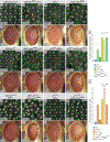
Figure 1:. Mask is required for patterning of the Drosophila eye independent of lattice cell number.
(A) Cartoon of a single ommatidium and surrounding lattice cells at 40 h APF. (B) Small region of a control retina at 40 h APF expressing lacZ, which does not disturb patterning. (C) Histograms of mean OMS scores for the genotypes in panels (B) and (D)-(G), ** denote p-value < 0.01. Retinas with (D) maskRNAi-v29541, (E) maskRNAi-v103411, (F) mask EY01848, or (G) mask RA. (B’) and (D’)-(G’) Tracings of images, with cone cells in orange, 1°s in yellow, lattice cells in green and bristle groups in grey. Confocal imaging settings were identical for panels (B) and (D). Images in (E-G) were generated with different imaging settings. See Table S3A,B for analyses of patterning defects. Representative eyes of (H) GMR>lacZ, (I) GMR>maskRNAi-v29541, (J) GMR>maskRNAi-v103411, (K) GMR>maskEY01848 and (L) GMR>mask RA adults. Retinas at 40 h APF expressing (M) lacZ or (N) rpr with the lattice-specific 54C-Gal4 driver, and (O) maskRNAi-v29541, (P) maskRNAi-v29541 and Diap1, (Q) lacZ (R) Diap1 and (S) maskEY01848 driven by GMR-Gal4. For (M)-(S) E-cad-detection was optimized during imaging to facilitate easier scoring of mis-patterning. 2 ommatidia are outlined (orange) to emphasize their shapes. Interommatidial cells are pseudo-colored green. Correctly-patterned 3°s are outlined in blue. Examples of grouped lattice cells are outlined in green and mis-oriented ommatidia indicated with yellow arrows. Abutting ommatidia are indicated with red lines. Yellow lines at 1°:1° boundaries emphasize relative size of 1° pairs.
Figure 2:
Figure 2:. Yki and Wts are required for eye morphogenesis.
Smalls regions of retinas at 40 h APF expressing (A) lacZ and Dcr-2, or (B) ykiRNAi and Dcr-2, (C) heterozygous for GMR-Gal4 or (D) expressing maskRNAi-v2954 and (E)-(H) representative eyes of adults of these genotypes. See Table S3D for further analyses of mis-patterning. Retinas at 40 h APF expressing (I) ectopic yki, and (J) ectopic yki and maskRNAi-v29541. (K) A retina heterozygous for GMR-Gal4 and ykib5, and (L) in addition with maskRNAi-v29541 expression. (M)-(P) Representative eyes of adults of genotypes (I)-(L). (Q) Mean OMS analyses at 40 h APF, for indicated genotypes. See Table S3E for further analyses of mis-patterning. (R) A retina heterozygous for GMR-Gal4 and a GMR-wts transgene, and (S) in addition with maskRNAi-v29541 expression. (T) A retina heterozygous for wtsX1 and GMR-Gal4 or (U) in addition maskRNAi-v29541. (V)-(Y) Representative eyes of adults of genotypes (R)-(U). (Z) Mean OMS analyses at 40 h APF, for indicated genotypes. See Table S3F for further analyses of mis-patterning. For panels (Q) and (Z), given the goal of testing modification of patterning in GMR>maskRNAi-v29541 retina when yki or wts expression was modified, significant changes in only these data are indicated. * denotes p-value < 0.1; ** denotes p-value < 0.01; ns = not significant. Abutting ommatidia are indicated with red lines; yellow * denote ommatidia missing 1°s; all other annotations as described in Figure 1. E-cad-imaging was optimized and images processed so that patterning defects could be scored.
Figure 3:
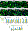
Figure 3:. AJs are not correctly organized when mask is reduced.
GMR>lacZ ommatidia at (A) 18, (B) 21, (C) 24 and (D) 27 h APF and (E)-(H) GMR>maskRNAi-v29541 ommatidia at analogous ages. Images (A)-(H) were gathered using identical confocal settings, but E-cad was enhanced in panels presented so that inconsistencies in AJ distribution can be observed. Orange arrows indicate examples of gaps in E-cad detection. (I) Cartoon of an ommatidium indicating the different AJs analyzed in (J), quantification of E-cad/AJ distribution in GMR>lacZ and GMR>maskRNAi-v29541 retinas. For N and p-values see Table S4B. (K) Quantification of amount of E-cad at AJs in GMR>lacZ and GMR> maskRNAi-v29541 retinas at 24 and 40 h APF. For N and p-values see Table S4C. In (J) and (K) all p-values were < 0.1 with the exception of the difference between E-cad coverage between 1° cells at 27 h APF (J). Error bars reflect standard error.
Figure 4:
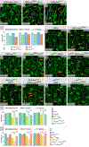
Figure 4:. AJ result from compromised Hippo pathway activity.
Ommatidia at 24 h APF expressing (A) lacZ and Dcr-2, (B) ykiRNAi and Dcr-2, or (C) ectopic wts and (D) quantification of AJ distribution; all p-values were < 0.1. For N and p-values see Table S4D. Ommatidia at 24 h APF heterozygous for (E) GMR-Gal4 or expressing (F) maskRNAi-v29541, (G) yki, and (H) yki and maskRNAi-v29541, heterozygous for (I) GMR-Gal4 and ykib5, or (J) in addition with maskRNAi-v29541 expression, heterozygous for (K) GMR-Gal4 and a GMR-wts transgene, and (L) in addition with maskRNAi-v29541 expression, heterozygous for (M) wtsX1 and GMR-Gal4, and (N) in addition maskRNAi-v29541. (O) Quantification of E-cad/AJ distribution for genotypes (E)-(J). For N and p-values see Table S4E. (P) Quantification of E-cad/AJ distribution for genotypes (E),(F),(K)-(N). For N and p-values see Table S4F. Given the goal of testing modification of AJ distribution by yki and wts in GMR>maskRNAi-v29541 retinas, significant changes in only these data are indicated in O and P. * denotes p-value < 0.1; ** denote p-value < 0.05; ns = not significant. Error bars reflect standard error. E-cad was enhanced in all images presented so that inconsistencies in AJ distribution can be observed. Orange arrows indicate examples of gaps in E-cad detection.
Figure 5:
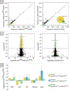
Figure 5:. Analyses of transcriptional changes in retinas in response to Mask.
(A) Scatterplots of gene expression (FKPM = fragments per kilobase per million reads) in retinas at 24 h APF in which mask expression was reduced (left) or increased (right), in comparison to control retinas. Yellow and green points indicate loci that were significantly differentially expressed when mask was modified (q < 0.05). Expression of 1674 genes was modified in GMR>maskRNAi-v29541 retinas and 255 in GMR>mask retinas, with 129 of these loci common to both data sets (see inset Venn diagram at right). (B) Volcano plots comparing significance (−log10 (q-value)) with the magnitude of expression change when mask expression was reduced (left) or increased (right) compared to corresponding controls. A q-value of 0.05 corresponds to −log10 (q-value) of ~1.3 and all yellow or green points indicate differentially-expressed loci. (C) Plot of FER, Vinc, Abi, Shroom, and wash expression assessed with RNA-seq and qRT-PCR in GMR>maskRNAi-v29541 or GMR>ykiRNAi retinas at 24 h APF. Error bars represent standard error.
Figure 6:
Figure 6:. FER mediates the role of Mask in regulating adhesion.
Ommatidia at 24 h APF (A) heterozygous for GMR-Gal4, (B) with ectopic FERP100, (C) maskRNAi-v29541 or, (D) FERP100 and maskRNAi-v29541. As before, tissue was imaged with identical confocal settings, but the images presented enhanced for better visualization of AJs. (E) Quantification of AJ (E-cad) distribution in retinas at 24 h APF. For N and p-values see Table S4G. (F)-(I) Retinas dissected at 40 h APF of genotypes (A)-(D). (J) Mean OMS values at 40 h APF, see Table S3H for detailed analyses. Given the goal of testing modification of GMR>maskRNAi-v29541 by FER, significant changes in only these data are indicated in (E) and (J); ** denotes p-value < 0.01; ns = not significant. Error bars reflect standard error. (K)-(N) Representative adult eyes of genotypes (A)-(D). (O) Ommatidia at 24 h APF heterozygous for GMR-Gal4 and FERX21, and (P) in addition with maskRNAi-v29541. (Q)-(R) Retinas of these two genotypes dissected at 40 h APF and (S)-(T) representative adult eyes. All annotations are as described in Figures 1–3.
Figure 7:
Figure 7:. Vinculin modifies the mis-patterning generated by maskRNAi.
Ommatidia at 24 h APF in retinas (A) heterozygous for GMR-Gal4, (B) with ectopic Vinc, (C) maskRNAi-v29541, and (D) with ectopic Vinc and maskRNAi-v29541. As before, eyes were imaged with identical confocal settings, but image panels enhanced for better visualization of AJs distribution. (E) AJ distribution at 24 h APF. For N and p-values see Table S4H. (F)-(I) Retinas dissected at 40 h APF of genotypes (A)-(D) and (J) mean OMS values at 40 h APF, see Table S3J for detailed analyses. Given the goal of testing modification of GMR>maskRNAi-v29541 by Vinc, significant changes in only these data are indicated in (E) and (J); * denotes p-value < 0.1; ** denotes p-value < 0.05; ns = not significant. Error bars reflect standard error. (K)-(N) representative adult eyes of genotypes (A)-(D). (O) Ommatidia at 24 h APF in retina heterozygous for GMR-Gal4 and Vinc Δ1, and (N) in addition maskRNAi-v29541 and (Q)-(R) retinas of these two genotypes dissected at 40 h APF and (S)-(T) representative adult eyes. Annotations are as described in Figures 1–3.

References

    1. Afgan E, Baker D, Batut B, van den Beek M, Bouvier D, Cech M, Chilton J, Clements D, Coraor N, Grüning BA, et al. (2018). The Galaxy platform for accessible, reproducible and collaborative biomedical analyses: 2018 update. Nucleic acids research 46, W537–W544. - PMC - PubMed
    1. Andrews S (2010). FastQC: a quality control tool for high throughput sequence data.
    1. Aragona M, Panciera T, Manfrin A, Giulitti S, Michielin F, Elvassore N, Dupont S, and Piccolo S (2013). A mechanical checkpoint controls multicellular growth through YAP/TAZ regulation by actin-processing factors. Cell 154, 1047–1059. - PubMed
    1. Bai H, Zhu Q, Surcel A, Luo T, Ren Y, Guan B, Liu Y, Wu N, Joseph NE, and Wang T-L (2016). Yes-Associated Protein impacts adherens junction assembly through regulating actin cytoskeleton organization. American Journal of Physiology-Gastrointestinal and Liver Physiology, ajpgi. 00027.02016. - PMC - PubMed
    1. Bao S, Fischbach K-F, Corbin V, and Cagan RL (2010). Preferential adhesion maintains separation of ommatidia in the Drosophila eye. Developmental Biology 344, 948–956. - PMC - PubMed

Publication types

MeSH terms

LinkOut - more resources