. 2020 May 25;9(5):1311.
doi: 10.3390/cells9051311.

Platelet Transforming Growth Factor-β1 Induces Liver Sinusoidal Endothelial Cells to Secrete Interleukin-6

Affiliations

Platelet Transforming Growth Factor-β1 Induces Liver Sinusoidal Endothelial Cells to Secrete Interleukin-6

Alexandre Balaphas et al. Cells. .

Abstract

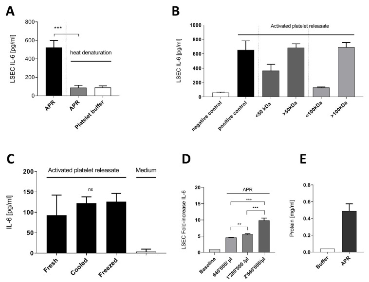
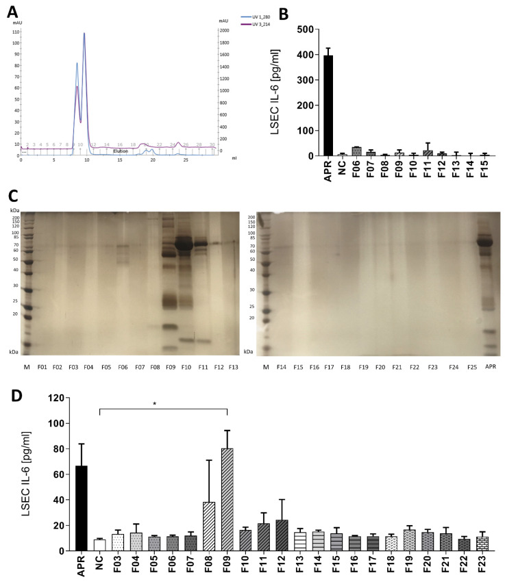
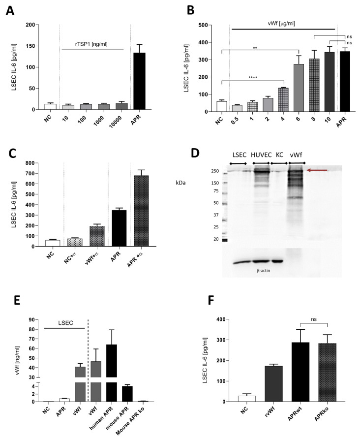
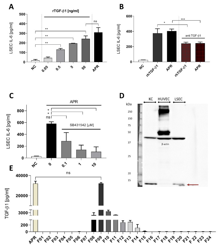
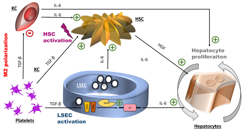
The roles and interactions of platelets and liver sinusoidal endothelial cells in liver regeneration are unclear, and the trigger that initiates hepatocyte proliferation is unknown. We aimed to identify the key factors released by activated platelets that induce liver sinusoidal endothelial cells to produce interleukin-6 (IL-6), a cytokine implicated in the early phase of liver regeneration. We characterized the releasate of activated platelets inducing the in vitro production of IL-6 by mouse liver sinusoidal endothelial cells and observed that the stimulating factor was a thermolabile protein. Following gel filtration, a single fraction of activated platelet releasate induced a maximal IL-6 secretion by liver sinusoidal endothelial cells (90.2 ± 13.9 versus control with buffer, 9.0 ± 0.8 pg/mL, p < 0.05). Mass spectroscopy analysis of this fraction, followed by in silico processing, resulted in a reduced list of 18 candidates. Several proteins from the list were tested, and only recombinant transforming growth factor β1 (TGF-β1) resulted in an increased IL-6 production up to 242.7 ± 30.5 pg/mL, which was comparable to non-fractionated platelet releasate effect. Using neutralizing anti-TGF-β1 antibody or a TGF-β1 receptor inhibitor, IL-6 production by liver sinusoidal endothelial cells was dramatically reduced. These results support a role of platelet TGF-β1 β1 in the priming phase of liver regeneration.

Keywords: Interleukin-6; hepatocytes; liver regeneration; livers sinusoidal cells; transforming growth factor β; von Willebrand factor.


Conflict of interest statement

S.B and A.T. are working for Regen Lab SA.

Figures

Figure 1
Figure 1
Characterization of activated platelet releasate soluble factor. (A): Mouse activated platelets releasate (APR) demonstrated lability to heat denaturation (95 °C, 15 min). (B): Using Amicon centrifugation filter of 50 kDa and 100 kDa cutoff, the supernatants containing proteins ˃ 50 kDa induced IL-6 secretion (C): Freshly prepared APR (Fresh) and after storage at 4 °C (Cooled) or at −20 °C (Frozen) still induced IL-6 secretion in LSEC. (D): Increasing platelet concentration in the preparation resulted in a significant fold increase of IL-6 production by LSEC. Baseline corresponds to basal IL-6 production by LSEC (E): Bradford assay estimating the total protein concentration of two mouse APR preparation of 640,000 platelet/µL. Buffer corresponds to platelet buffer. Presented data are representative of 2 or 3 independent experiments. *** p-value between 0.0001 and 0.001, ** p-value between 0.001 and 0.01 (t-test), ns: non-significant.
Figure 2
Figure 2
Immunofluorescence staining for interleukin-6 in liver sinusoidal endothelial cells and Kupffer cells. (A): Isolated liver sinusoidal endothelial cells (LSEC) were stained for stabilin-2 (green), and their nuclei were stained with Hoechst (blue). (B): In the absence of stimulation, IL-6 was not detected in LSEC (not shown). In contrast, after incubation of LSEC with APR, IL-6 (green) was detected in vesicles, with a homogeneous distribution in the cytoplasm. (C): Isolated Kupffer cells (KC) were stained for IBA-1 (red) and their nuclei were stained with Hoechst (blue). (D): KC were stimulated with lipopolysaccharide (0.5 mg/mL) and exhibited a different cytoplasmic distribution of IL-6 than LSEC. In LSEC, IL-6 was more homogeneously distributed in the cytoplasm (B) around the nuclei, whereas in KC, IL-6 appeared in clustered vesicles (D).
Figure 3
Figure 3
Gel filtration on a Superdex 75 increase column. (A): UV protein absorbance at 280 or 214 nm. All proteins of the sample were eluted from fraction 09 to 12. (B): Control experiment: eluted fractions of applied tyrode buffer and purified bovine thrombin were used for inducing IL-6 secretion by LSEC. Thrombin in tyrode buffer fractions did not activate LSEC. NC: negative control, tyrode buffer. (C): Silver-stained 12% polyacrylamide gel after SDS-PAGE showing proteins in APR fractions obtained after gel filtration. Proteins were mainly found in fractions 09 to 12. M: calibration, APR: non-fractionated APR. (D): Selected fractions were added to LSEC and IL-6 secretion assessed after 24 h of culture. Solely fraction 09 (F09) induced a statistically different IL-6 response compared to tyrode buffer, the negative control (NC), * p-value between 0.01 and 0.05 (t-test). APR prepared from 640,000 platelets/µL. n = 2. If not otherwise specified, presented data are representative of 2 or 3 independent experiments.
Figure 4
Figure 4
Characterization of soluble factors potentially inducing interleukin-6 secretion by liver sinusoidal endothelial cells. (A): Concentrations of thrombospondin 1 (TSP1) from 10 to 1000 ng/mL did not stimulate IL-6 secretion by LSEC. NC: negative control, tyrode buffer. (B): purified full-length human von Willebrand factor (vWF) was titrated on mouse LSEC. High concentrations of vWF resulted in an effect similar to human APR (positive control). NC: negative control, culture medium. (C): Neutralization of human APR or 8 μg/mL purified human vWF with anti-human vWF polyclonal antibody (α) 1/150. Combination of the antibody with APR or vWF resulted in an unexpected increase of IL-6 secretion by LSEC. NC: negative control, culture medium. (D): Western blot for expression of vWF in LSEC, HUVEC, KC as a positive control, human purified full-length vWF (2.5 μg). Contrarily, to HUVEC, LSEC and KC demonstrated low expression of vWF (250 kDa band, burgundy arrow). Purified full-length vWF exhibited several unspecific bands that were similar to HUVEC. Loading was controlled with β-actin. 7.5% acrylamide gel. n = 1 (E): Presence of mouse vWF was evaluated by ELISA in LSEC medium after treatment with APR. Human and mouse APR shows high amounts of vWF. NC: negative control, culture medium. (F): APR prepared from mouse double ko for vWF gene. APR was still able to induce IL-6 secretion by LSEC, 3 replicas. NC: negative control, culture medium. n = 1. **** p < 0.0001, ** p-value between 0.001, 0.01 (t-test),2 ns: non-significant. If not otherwise specified, presented data are representative of 2 or 3 independent experiments.
Figure 5
Figure 5
Transforming growth factor β1 is a strong candidate to activated platelet releasate soluble factor. (A): LSEC titration with recombinant human TGF-β1 induced a dose-response on IL-6 secretion, and at 50 ng/mL, there was no statistically significant difference with human APR. NC: negative control, culture medium. (B): Using anti-human TGF-β1, we were able to reduce the effect of APR or recombinant human TGF-β1 by 50%. Antibody concentration: 5 μg/mL, rTGF-β1 concentration: 5 ng/mL. NC: negative control, tyrode buffer. (C): TGF-β1 signaling blockade using the serine–threonine inhibitor SB431542 at increasing concentration. NC: negative control, DMSO (using DMSO with APR did not result in a decrease of IL-6 production). (D): Western blot for expression of TGF-β1 in KC, HUVEC and LSEC. TGF-β1 appeared in reduced condition as a monomer of 12.8 kDa (burgundy arrow) and was detected in KC and LSEC but not in HUVEC. Loading was controlled with β-actin. 10% acrylamide gel. n = 1. (E): TGF-β1 concentration in each fraction, obtained by ELISA, directly correlated with the IL-6 response pattern by LSEC. Indeed, fraction 09 showed a dramatic amount of TGF-β1 that were similar to the amounts found in non-fractionated human APR, n = 2. If not otherwise specified, presented data are representative of 2 or 3 independent experiments. *** p-value between 0.0001 and 0.001, ** p-value between 0.001 and 0.01, * p-value between 0.01 and 0.05 (t-test), ns: non-significant.
Figure 6
Figure 6
Potential effects of transforming growth factor β1 and interleukin-6 at the early stage of liver regeneration. The initial action of platelet TGF-β1 is the induction of the expression of the IL-6 gene by LSEC, probably through the canonical small mothers against decapentaplegic SMAD transcription factor complex. IL-6 promotes hepatocyte proliferation. Additionally, platelets TGF-β1 is connected to inflammation through its effects on hepatic stellate cells (HSC) (cell activation) and on Kuppfer cells (KC) (M2 polarization). HGF: hepatocyte growth factor, HSC: hepatic stellate cells, IL-6: interleukin-6, LSEC: liver sinusoidal endothelial cells, TGF- β1: transforming growth factor β1.

References

    1. Meyer J., Lejmi E., Fontana P., Morel P., Gonelle-Gispert C., Bühler L. A focus on the role of platelets in liver regeneration: Do platelet-endothelial cell interactions initiate the regenerative process? J. Hepatol. 2015;63:1263–1271. doi: 10.1016/j.jhep.2015.07.002. - DOI - PubMed
    1. Meyer J., Balaphas A., Fontana P., Sadoul K., Morel P., Gonelle-Gispert C., Bühler L. Platelets in liver regeneration. ISBT Sci. Ser. 2017;12:455–462. doi: 10.1111/voxs.12382. - DOI
    1. Balaphas A., Meyer J., Sadoul K., Fontana P., Morel P., Gonelle-Gispert C., Bühler L.H. Platelets and Platelet-Derived Extracellular Vesicles in Liver Physiology and Disease. Hepatol. Commun. 2019;3:855–866. doi: 10.1002/hep4.1358. - DOI - PMC - PubMed
    1. Kirschbaum M., Karimian G., Adelmeijer J., Giepmans B.N.G., Porte R.J., Lisman T. Horizontal RNA transfer mediates platelet-induced hepatocyte proliferation. Blood. 2015;126:798–806. doi: 10.1182/blood-2014-09-600312. - DOI - PubMed
    1. Peng Q., Yeh H., Wei L., Enjyoj K., Machaidze Z., Csizmad E., Schuetz C., Lee K.M., Deng S., Robson S.C., et al. Mechanisms of xenogeneic baboon platelet aggregation and phagocytosis by porcine liver sinusoidal endothelial cells. PLoS ONE. 2012;7:e47273. doi: 10.1371/journal.pone.0047273. - DOI - PMC - PubMed

LinkOut - more resources