. 2020 May 25;12(5):578.
doi: 10.3390/v12050578.

Isolation of a Chinook Salmon Bafinivirus (CSBV) in Imported Goldfish Carassius auratus L. in the United Kingdom and Evaluation of Its Virulence in Resident Fish Species

Affiliations

Isolation of a Chinook Salmon Bafinivirus (CSBV) in Imported Goldfish Carassius auratus L. in the United Kingdom and Evaluation of Its Virulence in Resident Fish Species

Irene Cano et al. Viruses. .

Abstract

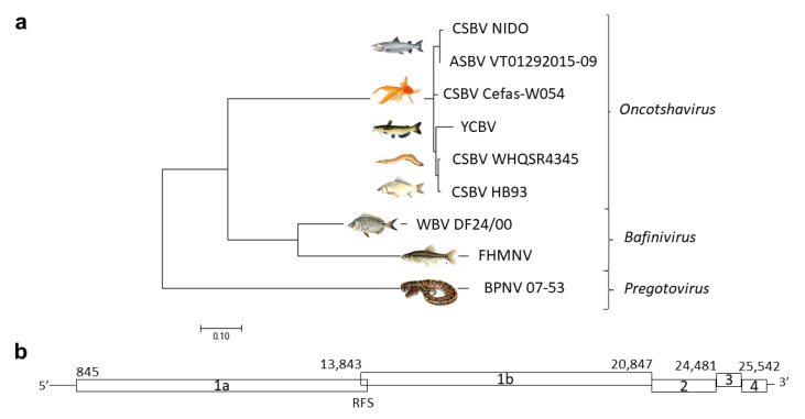
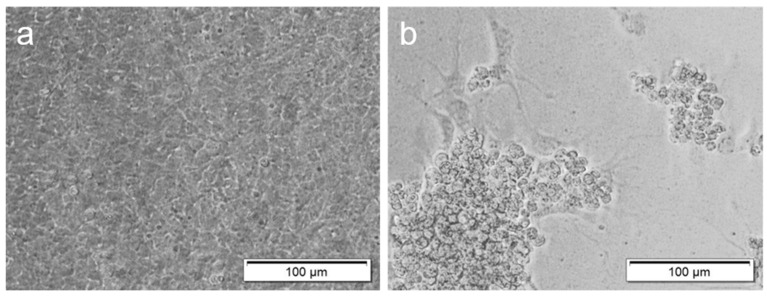
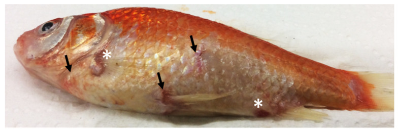
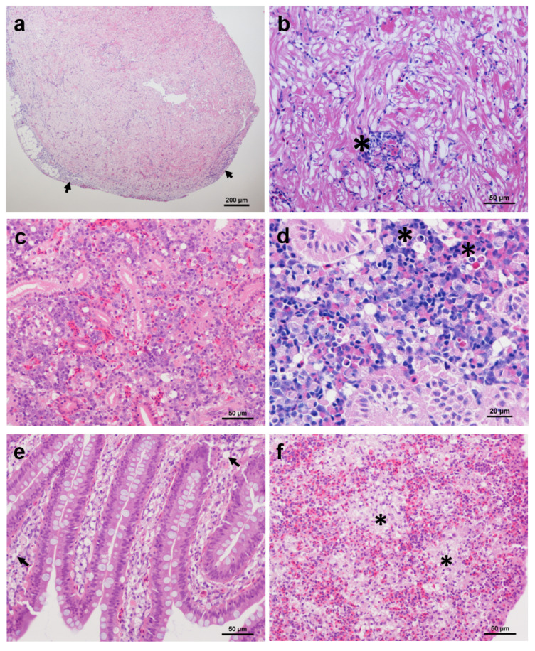
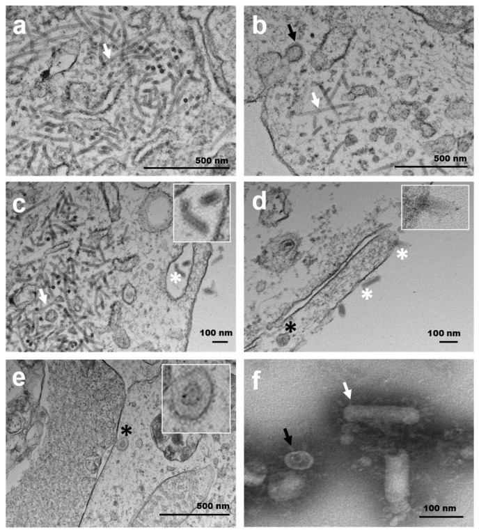
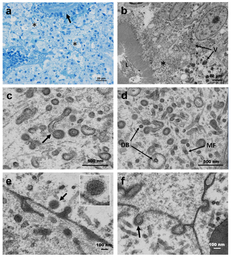
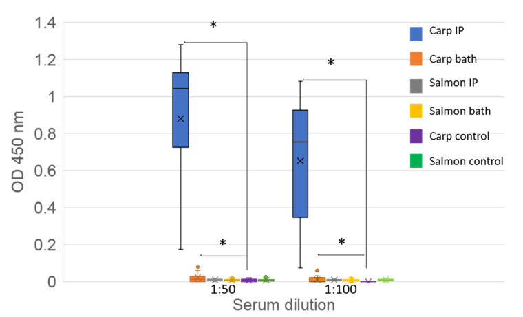
This is the first record of a fish nidovirus isolated from a consignment of goldfish at the United Kingdom (UK) border. The full-length viral genome was 25,985 nt, sharing a 97.9% nucleotide identity with the Chinook salmon bafinivirus (CSBV) NIDO with two deletions of 537 and 480 nt on the ORF Ia protein. To assess the potential impact on UK fish species, Atlantic salmon, common carp and goldfish were exposed to the virus via an intraperitoneal (IP) injection and bath challenge. Moribundity was recorded in only 8% of IP-injected goldfish. A high viral load, ≈107 of the CSBV PpIa gene, was measured in the kidney of moribund goldfish. Mild histopathological changes were observed in the kidneys of challenged carps. Ultrastructural observations in renal tubule epithelial cells of goldfish showed cylindrical tubes (≈15 nm in diameter) and tubular structures budding spherical virions (≈200 nm in diameter) with external spike-like structures. Negative staining showed both circular and bacilliform virions. Seroconversion was measured in common carp and goldfish but not in Atlantic salmon. This study reinforces the potential risk of novel and emerging pathogens being introduced to recipient countries via the international ornamental fish trade and the importance of regular full health screens at the border inspection posts to reduce this risk.

Keywords: border inspection post; common carp; diagnostics; emerging pathogen; goldfish; nidovirus.


Conflict of interest statement

The authors declare no conflict of interest.

Figures

Figure 1
Figure 1
(a) Neighbor-joining tree showing the phylogenetic relationship of the complete genome sequence of chinook salmon bafinivirus (CSBV) isolate Cefas-W054 (accession no. MT123520) and a selection of related tobaniviruses. GenBank accession: CSBV isolate NIDO (CSBV NIDO, no. KJ681496), Atlantic salmon bafinivirus isolate VT01292015-09 (ASBV VT01292015-09, no. KY130432), yellow catfish bafinivirus (YCBV, no. MH822145.1), CSBV isolate HB93 (MH171482), CSBV isolate WHQSR4345 (MG600027), white bream virus (WBV) isolate DF24/00 (DQ898157), fathead minnow nidovirus (FHMNV, no. GU002364) and ball python nidovirus (BPNV) isolate 07-53 (KJ541759). (b) CSBV Cefas-W054 predicted ORFs. Numbers indicate the predicted translation start codons. RFS: ribosomal frameshift in the ORF 1ab.
Figure 2
Figure 2
Cytopathic effects (CPEs) in fathead minnow (FHM) cells inoculated with goldfish chinook salmon bafinivirus Cefas-W054 at 7 days post-inoculation incubated at 20 °C. (a) FHM control cells. (b) FHM inoculated cells. CPEs consisted of detached cells (plaques) and rounded pyknotic and clumped cells.
Figure 3
Figure 3
Clinical signs of goldfish chinook salmon bafinivirus Cefas-W054 observed in an intraperitoneally injected goldfish, consisting of petechial haemorrhages in the skin and the base of the fins (arrows) and skin ulcerations (asterisks).
Figure 4
Figure 4
Histopathology associated with chinook salmon bafinivirus Cefas-W054. (a,b) Heart section of an intraperitoneally (IP)-injected goldfish showing (a) epicarditis (arrows) and (b) focal inflammation (asterisk); (c,d) Kidney section of an IP-injected goldfish (c) and a bath infected common carp (d) showing multifocal vacuolisation of haematopoietic tissue and cellular necrosis (asterisks); (e,f) IP-injected goldfish showing vacuolisation of the lamina propria (arrows) (e) and regions of vacuolated splenocytes (asterisks) (f). Haematoxylin and eosin staining was used.
Figure 5
Figure 5
Transmission electron micrographs of FHM (a,b) and E11 cells (cf) cells inoculated with chinook salmon bafinivirus Cefas-W054 isolate. (ac) Spherical (black arrows) and bacillary (white arrows) viral nucleocapsids were observed within the cytoplasm of these cells. (ce) Mature enveloped spherical (black asterisks) and bacillary virions (white asterisks). Top inserts show the detail of mature virions with spikes on the surface. (f) Transmission electron (negative staining) of purified virions from the E11 cell supernatant. The external surface of spikes can be seen surrounding spherical (black arrows) and rod-shaped (white arrows) virions.
Figure 6
Figure 6
(a) Semithin section of the kidney of moribund goldfish showing cellular necrosis in interstitial cells (asterisks) and renal tubule epithelium (arrow). (bf) Virogenesis in the proximal renal tubule epithelium. (b) Semithin resin section showing the apical region of the infected cell bordering the renal tubule lumen (L) and infected epithelial cell with numerous large vacuoles (V) containing granular and more electron-dense material that was mostly located between the nucleus and the viral assembly area (asterisk) in the apical portion of the cell. (c) Virions budding from tubular structures comprising the endoplasmic reticulum with the insertion of external microtubular material (arrow). (d) Internally fuzzy-coated vesicles with internal dense bodies (DB) and similar structures in the cytosol. The tubular structures are seen in (a,b); microfilaments (MF) and viral budding are also visible. (e,f) Viral budding from renal interstitial cell membranes. The top inserts show the detail of mature virions with spikes on the surface.
Figure 7
Figure 7
ELISA detection of CSBV antibodies in serum from challenged common carp and Atlantic salmon. Sera tested at 1:50 and 1:100 dilutions. Each bar represents from each group the median (cross), upper and lower quartile (box) and upper and lower extreme (line) of the optical density (OD) at 450 nm. Single points indicate outliers. Sample number per group: 15. IP: intraperitoneally injected fish; bath: bath infected fish. Asterisks (*) denotes significant differences (p < 0.5) between the treated and control groups.

References

    1. ORNAMENTAL AQUATIC TRADE ASSOCIATION EU Ornamental Fish Import & Export Statistics 2017 (Third Countries & Intra-EU Community Trade) [(accessed on 13 January 2020)]; Available online: https://www.ornamentalfish.org/wp-content/uploads/EU-Trade-Stats-Report-....
    1. Whittington R., Chong R. Global trade in ornamental fish from an Australian perspective: The case for revised import risk analysis and management strategies. Prev. Vet. Med. 2007;81:92–116. doi: 10.1016/j.prevetmed.2007.04.007. - DOI - PubMed
    1. Copp G.H., Wesley K.J., Vilizzi L. Pathways of ornamental and aquarium fish introductions into urban ponds of Epping Forest (London, England): The human vector*. J. Appl. Ichthyol. 2005;21:263–274. doi: 10.1111/j.1439-0426.2005.00673.x. - DOI
    1. Taylor N.G., Peeler E.J., Denham K., Crane C., Thrush M.A., Dixon P., Stone D.M., Way K., Oidtmann B. Spring viraemia of carp (SVC) in the UK: The road to freedom. Prev. Vet. Med. 2013;111:156–164. doi: 10.1016/j.prevetmed.2013.03.004. - DOI - PubMed
    1. Taylor N.G., Norman R., Way K., Peeler E.J. Modelling the koi herpesvirus (KHV) epidemic highlights the importance of active surveillance within a national control policy. J. Appl. Ecol. 2011;48:348–355. doi: 10.1111/j.1365-2664.2010.01926.x. - DOI

Publication types