Review
. 2020 May 26;25(11):2471.
doi: 10.3390/molecules25112471.

Collective Locomotion of Human Cells, Wound Healing and Their Control by Extracts and Isolated Compounds from Marine Invertebrates

Affiliations
Review

Collective Locomotion of Human Cells, Wound Healing and Their Control by Extracts and Isolated Compounds from Marine Invertebrates

Claudio Luparello et al. Molecules. .

Abstract

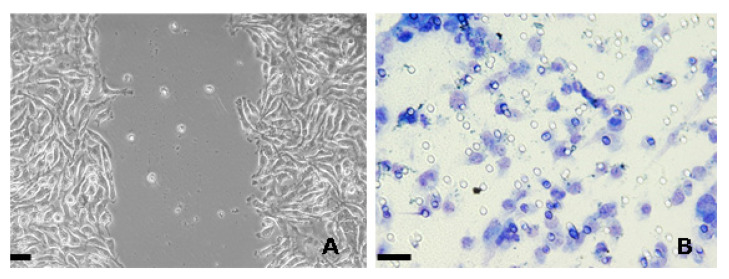
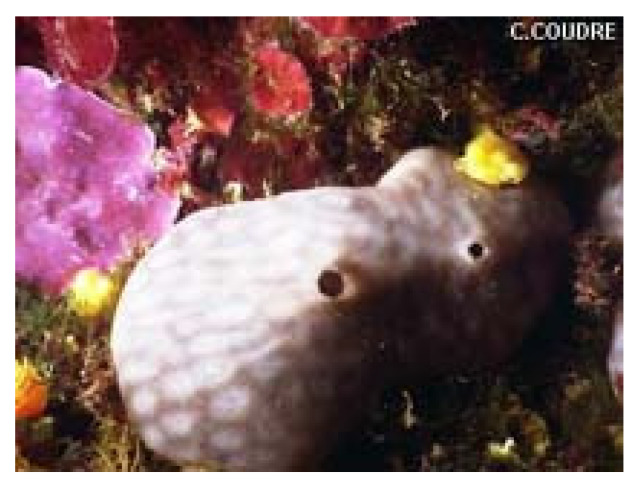
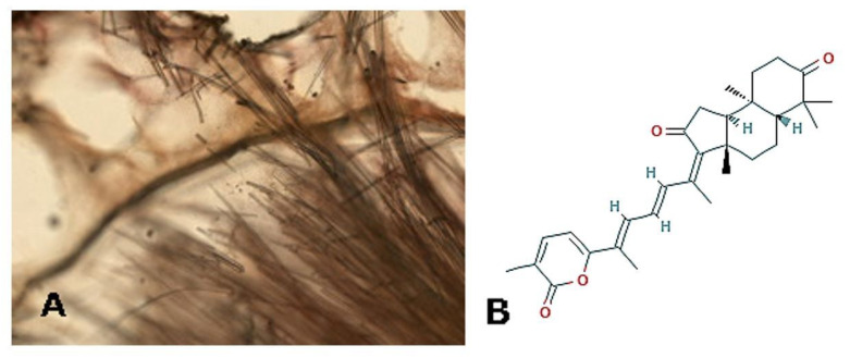
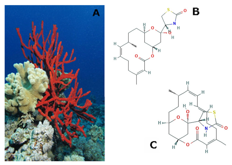
The collective migration of cells is a complex integrated process that represents a common theme joining morphogenesis, tissue regeneration, and tumor biology. It is known that a remarkable amount of secondary metabolites produced by aquatic invertebrates displays active pharmacological properties against a variety of diseases. The aim of this review is to pick up selected studies that report the extraction and identification of crude extracts or isolated compounds that exert a modulatory effect on collective cell locomotion and/or skin tissue reconstitution and recapitulate the molecular, biochemical, and/or physiological aspects, where available, which are associated to the substances under examination, grouping the producing species according to their taxonomic hierarchy. Taken all of the collected data into account, marine invertebrates emerge as a still poorly-exploited valuable resource of natural products that may significantly improve the process of skin regeneration and restrain tumor cell migration, as documented by in vitro and in vivo studies. Therefore, the identification of the most promising invertebrate-derived extracts/molecules for the utilization as new targets for biomedical translation merits further and more detailed investigations.

Keywords: cell migration; marine invertebrates; wound healing.


Conflict of interest statement

The authors declare no conflict of interest.

Figures

Figure 1
Figure 1
(A) Phase-contrast microscopic image of a scratch created in a monolayer of HEPG2 liver carcinoma cells. (B) Light microscopic image of MDA-MB231 breast carcinoma cells migrated through the pores of a polycarbonate filter in a Boyden chamber assay, fixed with 95% ethanol, and stained with 0.02 g toluidine blue/mL. Bar = 10 μm.
Figure 2
Figure 2
A specimen of Chondrosia reniformis demosponge. © Bernard Picton (CC BY-NC-SA 4.0); http://www.marinespecies.org/.
Figure 3
Figure 3
(A) Image of a paraffin section of Jaspis stellifera demosponge. Photo by D. Drew (YPM IZ 087028). Courtesy of the Peabody Museum of Natural History, Division of Vertebrate Paleontology, Yale University; peabody.yale.edu. (B) 2D structure of stellettin B; https://pubchem.ncbi.nlm.nih.gov/compound/5352082#section=2D-Structure.
Figure 4
Figure 4
(A) A specimen of Negombata magnifica demosponge. Photo © Alexander Vasenin (CC BY-SA 3.0) https://en.wikipedia.org/. (B) Two-dimensional (2D) structure of latrunculin B; https://pubchem.ncbi.nlm.nih.gov/compound/Latrunculin-B#section=2D-Structure. (C) 2D structure of latrunculin A; https://pubchem.ncbi.nlm.nih.gov/compound/Latrunculin-a#section=2D-Structure.
Figure 5
Figure 5
(A) A specimen of Pseudoceratina arabica demosponge © Danièle Heitz; https://nomadica.jimdofree.com/. (B) 2D structure of moloka’iamine; https://pubchem.ncbi.nlm.nih.gov/compound/Moloka_Iamine#section=Structures. (C) 2D structure of hydroxymoloka’iamine; https://pubchem.ncbi.nlm.nih.gov/compound/25016152. (D) A specimen of Suberea mollis demosponge; Abbas et al., 2014 (CC BY 3.0).
Figure 6
Figure 6
(A) A specimen of Siphonocalina siphonella demosponge © Alexander Vasenin (CC BY-SA 3.0); https://commons.wikimedia.org/. (B) 2D structure of sipholenol A; https://pubchem.ncbi.nlm.nih.gov/compound/Sipholenol-A#section=Structures.
Figure 7
Figure 7
Examples of specimens of Xestospongia sp. (A) X. muta © Renaud Houdinet; https://flickr.com/; (B) X. testudinaria, photo by Albert Kok (Public domain) https://commons.wikimedia.org/.
Figure 8
Figure 8
(A) A specimen of Cladiella australis soft coral © David (CC BY-NC-SA 4.0) https://www.marinespecies.org/. (B) 2D structure of austrasulfone; https://pubchem.ncbi.nlm.nih.gov/compound/50907657#section=Structures.
Figure 9
Figure 9
(A) A specimen of Cladiella pachyclados soft coral © Danièle Heitz; https://nomadica.jimdofree.com/. (B) Two-dimensional (2D) structure of pachycladin A; https://pubchem.ncbi.nlm.nih.gov/compound/Pachycladin-A#section=2D-Structure. (C) 2D structure of sclerophytin F methyl ether; https://pubchem.ncbi.nlm.nih.govcompound/21635655. (D) 2D structure of polyanthelin A; https://pubchem.ncbi.nlm.nih.gov/compound/12168107. (E) 2D structure of sclerophytin A; https://pubchem.ncbi.nlm.nih.gov/compound/Sclerophytin-A#section=2D-Structure.
Figure 10
Figure 10
(A) A specimen of Sarcophyton crassocaule soft coral © Leen Van Ofwegen (CC BY-NC-SA 4.0); https://www.marinespecies.org/ (B) 2D structure of 13-acetoxysarcocrassolide; https://pubchem.ncbi.nlm.nih.gov/compound/21775908.
Figure 11
Figure 11
(A) 2D structure of 11-dehydrosinulariolide; http://www.chemspider.com/Chemical-Structure.10470882.html. (B) 2D structure of sinularin; https://pubchem.ncbi.nlm.nih.gov/compound/Sinularin#section=2D-Structure.
Figure 12
Figure 12
An image of Clavularia koellikeri; Wang et al. (2019) [87].
Figure 13
Figure 13
An indivividual of Cyanea capillata © Alexander Semenov; https://www.flickr.com/.
Figure 14
Figure 14
A specimen of Rhopilema esculentum. © Bill Abbott (CC BY-SA 2.0); https://en.wikipedia.org/.
Figure 15
Figure 15
(A) A specimen of Uroteuthis singhalensis; http://www.borneomolluscs.com. (B) 2D structure of astaxanthin; https://pubchem.ncbi.nlm.nih.gov/compound/Astaxanthin#section=2D-Structure.
Figure 16
Figure 16
A specimen of Sepia kobiensis. © Maynard Hogg (CC BY-NC-SA 2.0); https://www.flickr.com/.
Figure 17
Figure 17
A specimen of Sepia officinalis. © Hans Hillewaert (CC BY-SA 4.0); http://www.marinespecies.org/.
Figure 18
Figure 18
(A) A group of Mytilus galloprovincialis bivalves. © Pillon Robert (CC BY-NC-SA 4.0); http://www.marinespecies.org/. (B) Images of the shell of Rapana venosa. © H. Zell (CC BY-SA 3.0); https://en.wikipedia.org/. (C) 2D structure of eicosapentaenoic acid; https://pubchem.ncbi.nlm.nih.gov/compound/Eicosapentaenoic-acid#section=2D-Structure. (D) 2D structure of docosahexenoic acid; https://pubchem.ncbi.nlm.nih.gov/compound/Docosahexaenoic-acid#section=Structures. (E) 2D structure of arachidonic acid; https://pubchem.ncbi.nlm.nih.gov/compound/Arachidonic-acid#section=2D-Structure.
Figure 19
Figure 19
Images of the shell of Pinctada imbricata. © Natural History Museum Rotterdam (CC BY-NC-SA 4.0); http://www.marinespecies.org/.
Figure 20
Figure 20
Images of the shell of Euchelus asper. © H. Zell (CC BY-SA 3.0); https://commons.wikimedia.org.
Figure 21
Figure 21
Images of the shell of Haliotis diversicolor squamata. © Natural History Museum Rotterdam (CC BY-NC-SA 4.0); http://www.marinespecies.org/.
Figure 22
Figure 22
Specimens of Euphausia superba. © Brett Wilks; http://www.antarctica.gov.au/.
Figure 23
Figure 23
An individual of Astropecten indicus. © Ria Tan (CC BY-NC-ND 2.0); https://www.flickr.com/.
Figure 24
Figure 24
(A) An individual of Himerometra magnipinna. © Andrew Green; https://reeflifesurvey.com/. (B) 2D structure of rhodoptilometrin; https://pubchem.ncbi.nlm.nih.gov/compound/Rhodoptilometrin#section=2D-Structure.
Figure 25
Figure 25
A specimen of Stichopus japonicus. © Alexander Semenov; https://www.flickr.com.
Figure 26
Figure 26
A specimen of Isostichopus badionotus. © Dan Hershman (CC BY 2.0); https://commons.wikimedia.org/.
Figure 27
Figure 27
An individual of Ophiocoma erinaceus. © Patrick Randall (CC BY-NC-SA 2.0); https://www.flickr.com/.
Figure 28
Figure 28
A specimen of Stichopus herrmanni. © Andrew Green; https://reeflifesurvey.com/.
Figure 29
Figure 29
(A) An image of Phallusia arabica. © Frédéric André; https://doris.ffessm.fr/. (B) 2D structure of n-hexadecanoic acid; https://pubchem.ncbi.nlm.nih.gov/compound/Palmitic-acid#section=2D-Structure.
Figure 30
Figure 30
An image of Phallusia nigra. © Rosana M. Rocha; http://invasions.si.edu/.
Figure 31
Figure 31
An image of Styela clava. © Claude Nozères (CC BY-NC-SA 4.0); http://www.marinespecies.org/.

Similar articles

Cited by

References

    1. Li L., He Y., Zhao M., Jiang J. Collective cell migration: Implications for wound healing and cancer invasion. Burns Trauma. 2013;1:21–26. doi: 10.4103/2321-3868.113331. - DOI - PMC - PubMed
    1. Camley B.A., Rappel W.-J. Physical models of collective cell motility: From cell to tissue. J. Phys. D Appl. Phys. 2017;50:113002. doi: 10.1088/1361-6463/aa56fe. - DOI - PMC - PubMed
    1. Ladoux B., Mège R.-M. Mechanobiology of collective cell behaviours. Nat. Rev. Mol. Cell Biol. 2017;18:743–757. doi: 10.1038/nrm.2017.98. - DOI - PubMed
    1. Eelen G., de Zeeuw P., Simons M., Carmeliet P. Endothelial Cell Metabolism in Normal and Diseased Vasculature. Circ. Res. 2015;116:1231–1244. doi: 10.1161/CIRCRESAHA.116.302855. - DOI - PMC - PubMed
    1. Potente M., Gerhardt H., Carmeliet P. Basic and Therapeutic Aspects of Angiogenesis. Cell. 2011;146:873–887. doi: 10.1016/j.cell.2011.08.039. - DOI - PubMed

LinkOut - more resources