Review
. 2020 May 26;9(6):1330.
doi: 10.3390/cells9061330.

Crucial Role of Lamin A/C in the Migration and Differentiation of MSCs in Bone

Affiliations
Review

Crucial Role of Lamin A/C in the Migration and Differentiation of MSCs in Bone

Natividad Alcorta-Sevillano et al. Cells. .

Abstract

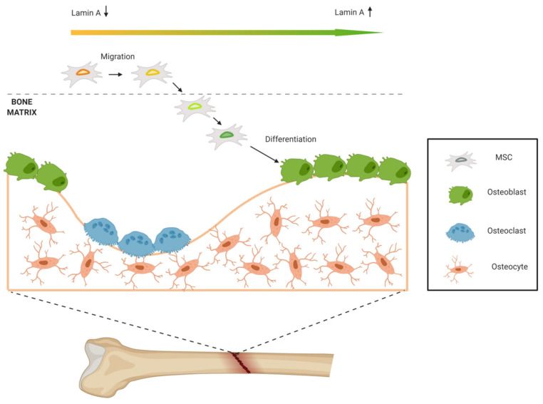
Lamin A/C, intermediate filament proteins from the nuclear lamina encoded by the LMNA gene, play a central role in mediating the mechanosignaling of cytoskeletal forces into nucleus. In fact, this mechanotransduction process is essential to ensure the proper functioning of other tasks also mediated by lamin A/C: the structural support of the nucleus and the regulation of gene expression. In this way, lamin A/C is fundamental for the migration and differentiation of mesenchymal stem cells (MSCs), the progenitors of osteoblasts, thus affecting bone homeostasis. Bone formation is a complex process regulated by chemical and mechanical cues, coming from the surrounding extracellular matrix. MSCs respond to signals modulating the expression levels of lamin A/C, and therefore, adapting their nuclear shape and stiffness. To promote cell migration, MSCs need soft nuclei with low lamin A content. Conversely, during osteogenic differentiation, lamin A/C levels are known to be increased. Several LMNA mutations present a negative impact in the migration and osteogenesis of MSCs, affecting bone tissue homeostasis and leading to pathological conditions. This review aims to describe these concepts by discussing the latest state-of-the-art in this exciting area, focusing on the relationship between lamin A/C in MSCs' function and bone tissue from both, health and pathological points of view.

Keywords: bone disease; bone formation; differentiation; lamin A/C; mechanosensing; mesenchymal stem cells (MSCs); migration.


Conflict of interest statement

The authors declare no conflict of interest. The funders had no role in the collection, analyses or interpretation of data; in the writing of the manuscript, or in the decision to publish the review.

Figures

Figure 1
Figure 1
Linker of Nucleus and Cytoskeleton (LINC) complex. This structure consists of SUN (Sad1 and UNC-84) proteins anchored in the inner nuclear membrane and nesprins anchored in the outer nuclear membrane. While nesprins interact with different proteins of the cytoskeleton such as actin, SUN proteins are linked with lamins that surround the nuclear envelope.
Figure 2
Figure 2
Mesenchymal stem cells’ (MSCs) fate is regulated by lamin A and vascular endothelial growth factor (VEGF) interaction. High levels of lamin A induce the expression of VEGF, which in turn induces the Runt-related transcription factor (RUNX2) transcription factor synthesis, responsible for osteoblast differentiation, inhibiting the adipocyte differentiation. However, low levels of VEGF lead to activation of the peroxisome proliferator-activated receptor (PPAR)-γ transcription factor, responsible of adipogenesis. Dashed lines represent the routes that are downregulated or compromised.
Figure 3
Figure 3
MSCs’ migration and differentiation. Lamin A/C levels in MSCs vary according to the microenvironment. Low lamin A/C levels are needed for migration, while high levels are required for osteogenic differentiation.
Figure 4
Figure 4
Chemical and mechanical factors that regulate MSCs’ migration and differentiation. These factors regulate lamin A/C levels, thus affecting nuclear shape and stiffness. In this way, MSCs’ migration and/or differentiation into osteogenic lineage depend on lamin A/C expression.

Similar articles

Cited by

References

    1. Burke B., Stewart C.L. The nuclear lamins: Flexibility in function. Nat. Rev. Mol. Cell. Biol. 2013;14:13–24. doi: 10.1038/nrm3488. - DOI - PubMed
    1. Beck L.A., Hosick T.J., Sinensky M. Isoprenylation is required for the processing of the lamin A precursor. J. Cell. Biol. 1990;110:1489–1499. doi: 10.1083/jcb.110.5.1489. - DOI - PMC - PubMed
    1. Holtz D., Tanaka R.A., Hartwig J., McKeon F. The CaaX motif of lamin A functions in conjunction with the nuclear localization signal to target assembly to the nuclear envelope. Cell. 1989;59:969–977. doi: 10.1016/0092-8674(89)90753-8. - DOI - PubMed
    1. Fong L.G., Ng J.K., Lammerding J., Vickers T.A., Meta M., Cote N., Gavino B., Qiao X., Chang S.Y., Young S.R., et al. Prelamin A and lamin A appear to be dispensable in the nuclear lamina. J. Clin. Investig. 2006;116:743–752. doi: 10.1172/JCI27125. - DOI - PMC - PubMed
    1. Shimi T., Pfleghaar K., Kojima S., Pack C.G., Solovei I., Goldman A.E., Adam S.A., Shumaker D.K., Kinjo M., Cremer T., et al. The A- and B-type nuclear lamin networks: Microdomains involved in chromatin organization and transcription. Genes Dev. 2008;22:3409–3421. doi: 10.1101/gad.1735208. - DOI - PMC - PubMed

Publication types

LinkOut - more resources