

Inertial Sensors as a Tool for Diagnosing Discopathy Lumbosacral Pathologic Gait: A Preliminary Research
- PMID: 32466525
- PMCID: PMC7345098
- DOI: 10.3390/diagnostics10060342
Inertial Sensors as a Tool for Diagnosing Discopathy Lumbosacral Pathologic Gait: A Preliminary Research
Abstract
Background: the goal of the study is to ascertain the influence of discopathy in the lumbosacral (L-S) segment on the gait parameters. The inertial sensors are used to determine the pathologic parameters of gait.
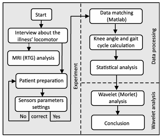
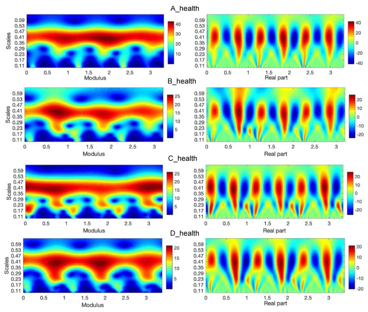
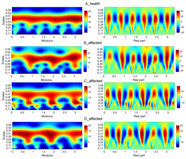
Methods: the study involved four patients (44, 46, 42, and 38 years). First, the goal of the survey was to analyze by a noninvasive medical test magnetic resonance imaging (MRI) of each patient. Next, by using inertial sensors, the flexion-extension of joint angles of the left and right knees were calculated. The statistical analysis was performed. The wavelet transform was applied to analyze periodic information in the acceleration data.
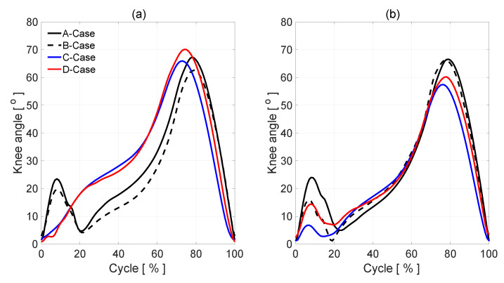
Results: in the patients with discopathy, the amount of knee flexion attained during stance phase is significantly lower than that of normal (health side), which could indicate poor eccentric control or a pain avoidance mechanism. The biggest differences are observed in the Initial Swing phase. Bending of the lower limb in the knee joint at this stage reaches maximum values during the entire gait cycle.
Conclusions: It has been difficult to quantify the knee angle during gait by visual inspection. The inertial measurement unit (IMU) system can be useful in determining the level of spine damage and its degree. In patients in the first stages of the intervertebral disc disease who may undergo conservative treatment, it may also partially delay or completely exclude the decision to perform a complicated imaging examination which is MRI, often showing a false positive result in this phase of the disease.
Keywords: computer modelling; human gait; inertial sensors; lumbar discopathy; wavelet analysis.
Conflict of interest statement
The authors declare no conflicts of interest.
Figures


References
-
- Glowinski S., Blazejewski A., Krzyzynski T. Inertial sensors and wavelets analysis as a tool for pathological gait identification. Adv. Intell. Syst. Comput. 2017;526:104–114. doi: 10.1007/978-3-319-47154-9_13. - DOI
LinkOut - more resources
Full Text Sources
