. 2020 May 26;21(11):3756.
doi: 10.3390/ijms21113756.

Nicotinamide Mononucleotide Administration Prevents Experimental Diabetes-Induced Cognitive Impairment and Loss of Hippocampal Neurons

Affiliations

Nicotinamide Mononucleotide Administration Prevents Experimental Diabetes-Induced Cognitive Impairment and Loss of Hippocampal Neurons

Krish Chandrasekaran et al. Int J Mol Sci. .

Abstract

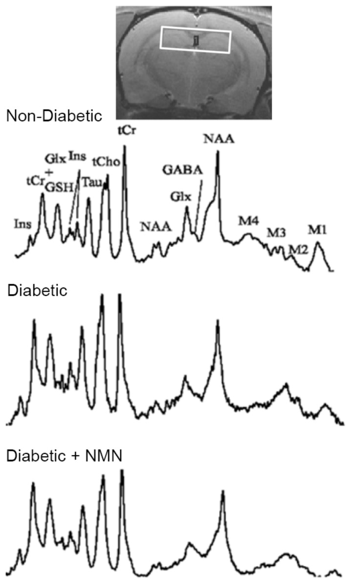
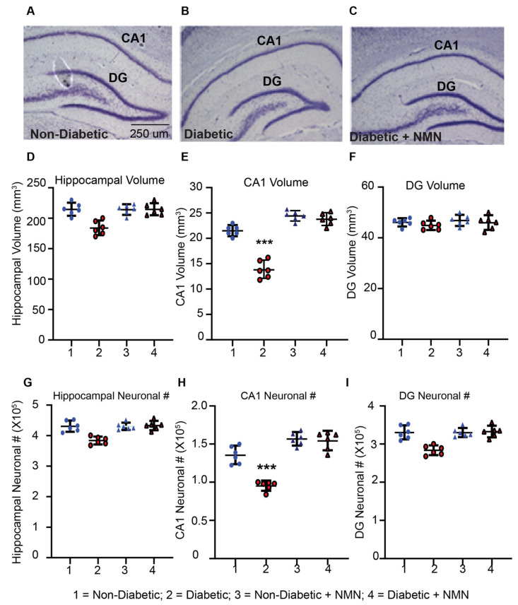
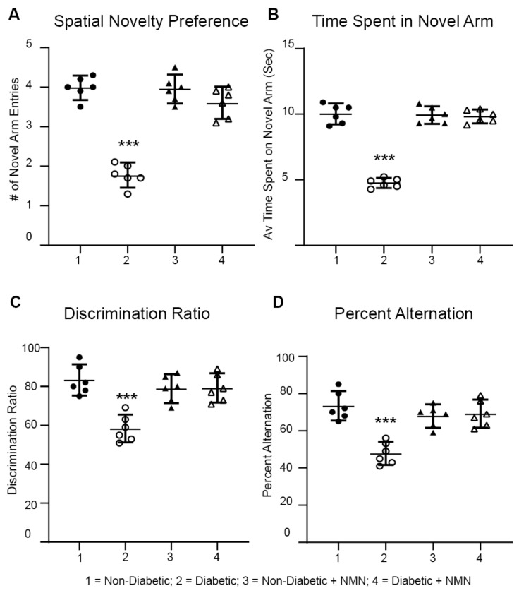
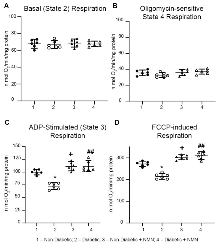
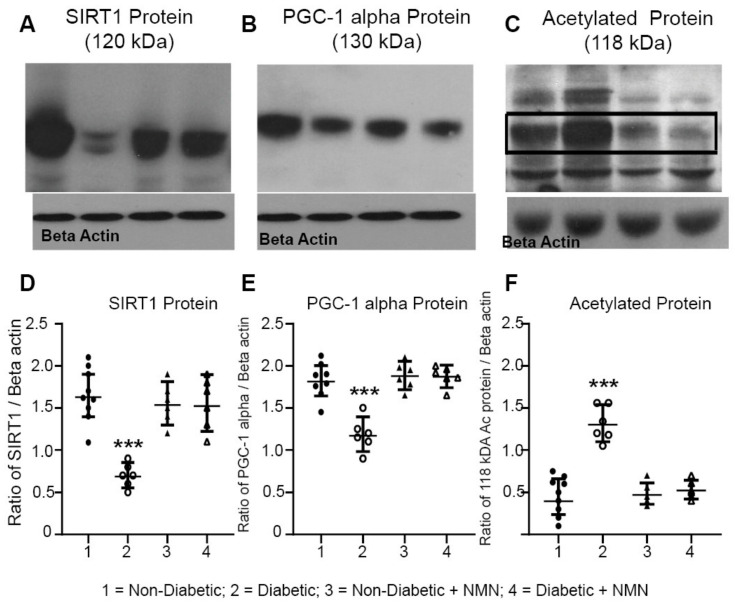
Diabetes predisposes to cognitive decline leading to dementia and is associated with decreased brain NAD+ levels. This has triggered an intense interest in boosting nicotinamide adenine dinucleotide (NAD+) levels to prevent dementia. We tested if the administration of the precursor of NAD+, nicotinamide mononucleotide (NMN), can prevent diabetes-induced memory deficits. Diabetes was induced in Sprague-Dawley rats by the administration of streptozotocin (STZ). After 3 months of diabetes, hippocampal NAD+ levels were decreased (p = 0.011). In vivo localized high-resolution proton magnetic resonance spectroscopy (MRS) of the hippocampus showed an increase in the levels of glucose (p < 0.001), glutamate (p < 0.001), gamma aminobutyric acid (p = 0.018), myo-inositol (p = 0.018), and taurine (p < 0.001) and decreased levels of N-acetyl aspartate (p = 0.002) and glutathione (p < 0.001). There was a significant decrease in hippocampal CA1 neuronal volume (p < 0.001) and neuronal number (p < 0.001) in the Diabetic rats. Diabetic rats showed hippocampal related memory deficits. Intraperitoneal NMN (100 mg/kg) was given after induction and confirmation of diabetes and was provided on alternate days for 3 months. NMN increased brain NAD+ levels, normalized the levels of glutamate, taurine, N-acetyl aspartate (NAA), and glutathione. NMN-treatment prevented the loss of CA1 neurons and rescued the memory deficits despite having no significant effect on hyperglycemic or lipidemic control. In hippocampal protein extracts from Diabetic rats, SIRT1 and PGC-1α protein levels were decreased, and acetylation of proteins increased. NMN treatment prevented the diabetes-induced decrease in both SIRT1 and PGC-1α and promoted deacetylation of proteins. Our results indicate that NMN increased brain NAD+, activated the SIRT1 pathway, preserved mitochondrial oxidative phosphorylation (OXPHOS) function, prevented neuronal loss, and preserved cognition in Diabetic rats.

Keywords: NAD+; NEDD4-1; NMN; PGC-1α; SIRT1; cognitive impairment; dementia; diabetes; mitochondria.


Conflict of interest statement

The authors declare no conflict of interest.

Figures

Figure 1
Figure 1
Example of the magnetic resonance spectroscopy spectra acquired at 3 months from Non-Diabetic, Diabetic, Diabetic + NMN, and Non-Diabetic + NMN rats. Only the biochemicals measurable spectra (15%) are reported. GABA = gamma amino butyric acid; Glu = glutamate; Gln = glutamine; PCh = phosphatidyl choline; NAA = N-acetyl aspartate; GSH = glutathione; NAAG = N-acetyl aspartyl glutamate. The data acquired are reported in Table 2.
Figure 2
Figure 2
Diabetes-induced loss of hippocampal volume and neuronal counts in CA1 after 3 months of diabetes is prevented by NMN-treatment. (AC) Coomassie-blue stained hippocampal section of Non-Diabetic, Diabetic, and Diabetic + NMN rat brains are shown. There was no difference in staining pattern between Non-Diabetic and Non-Diabetic + NMN rats. (D) Comparison of the total hippocampal volume between Non-Diabetic and Diabetic rats showed no significant difference. (E) Comparison of the CA1 hippocampal neuronal volume between Non-Diabetic and Diabetic rats showed a significant difference (p < 0.05) using a one-way ANOVA test with a post hoc Tukey test but no significant difference between hippocampal neuronal volume in Non-Diabetic and Diabetic + NMN rats. (F) Comparison of the dentate gyrus (DG) volume between Non-Diabetic and Diabetic rats showed no significant difference between the groups. (G) Comparison of the total hippocampal neuronal number (#) between Non-Diabetic and Diabetic rats showed no significant difference. (H) Comparison of the CA1 hippocampal neuronal number (#) between Non-Diabetic and Diabetic rats showed a significant difference (p < 0.05) using one-way ANOVA test with a post hoc Tukey test but no significant difference between Non-Diabetic and Diabetic + NMN rats. (I) Comparison of the DG neuronal number (#) between Non-Diabetic and Diabetic rats showed no significant difference among the groups. *** p < 0.001 Diabetes vs. Diabetes + NMN.
Figure 3
Figure 3
Impaired memory in Diabetic rats was prevented by NMN treatment. Diabetic rats displayed a reduced spontaneous alternation and novel spatial preference compared to those of Non-Diabetic rats at 3 months. (AC) Spontaneous spatial novelty preference test: NMN prevents a diabetes-induced decrease in the number (#) of novel arm entries (A), the average time spent in the novel arm (B), and the discrimination ratio (C). (D) Spontaneous alternation test: NMN prevents a diabetes-induced decrease in the spontaneous alternation %. (N = 6). *** p < 0.001.
Figure 4
Figure 4
Impaired mitochondrial respiration in Diabetic rats was prevented by NMN treatment. Oxygen consumption rate (OCR) was measured at basal level with the subsequent and sequential addition of ADP, oligomycin, FCCP, and rotenone + antimycin A to hippocampal mitochondria. State 2, State 3, State 4 (oligomycin-sensitive), and FCCP-induced respiration rates were measured. ADP-stimulated and uncoupled respiration was significantly decreased in Diabetic rats compared to Non-Diabetic rats. Administration of NMN significantly increased both ADP-stimulated and uncoupled respiration. Spare respiratory capacity was calculated after subtracting the basal respiration from uncoupled respiration and was significantly higher (p < 0.05) in NMN treated Non-Diabetic and Diabetic hippocampal mitochondria. * p < 0.05 Diabetic vs. Non-Diabetic; + p < 0.05 Non-Diabetic vs. Non-Diabetic + NMN; ## p < 0.01 Diabetic vs. Diabetic + NMN.
Figure 5
Figure 5
(AC) Western blot analysis of NAD+-consuming enzyme SIRT1 and acetylated proteins. Hippocampal protein extracts were prepared from Non-Diabetic, Diabetic, Non-Diabetic + NMN, and Diabetic + NMN rat brains. Preparation of hippocampal protein extracts, blot analysis, the source, and the dilution of the antibodies used are described in Section 4. (DF) Quantification of the intensity of the bands are shown. Significant decrease in SIRT1 protein and PGC-1α protein levels, and an increase in acetylated 118 kDa protein were observed in Diabetic samples, but not in NMN-treated Diabetic + NMN samples. *** p < 0.001 Diabetic vs. Diabetic + NMN.

References

    1. Grunblatt E., Bartl J., Riederer P. The link between iron, metabolic syndrome, and Alzheimer’s disease. J. Neural Transm. 2011;118:371–379. doi: 10.1007/s00702-010-0426-3. - DOI - PubMed
    1. Ott A., Stolk R.P., van Harskamp F., Pols H.A., Hofman A., Breteler M.M. Diabetes mellitus and the risk of dementia: The Rotterdam Study. Neurology. 1999;53:1937–1942. doi: 10.1212/WNL.53.9.1937. - DOI - PubMed
    1. Ma X., Sun Z., Han X., Li S., Jiang X., Chen S., Zhang J., Lu H. Neuroprotective effect of resveratrol via activation of Sirt1 signaling in a rat model of combined diabetes and Alzheimer’s disease. Front. Neurosci. 2019;13:1400. doi: 10.3389/fnins.2019.01400. - DOI - PMC - PubMed
    1. Zhou Y., Lian S., Zhang J., Lin D., Huang C., Liu L., Chen Z. Mitochondrial perturbation contributing to Cognitive Decline in Streptozotocin-Induced Type 1 Diabetic Rats. Cell Physiol. Biochem. 2018;46:1668–1682. doi: 10.1159/000489243. - DOI - PubMed
    1. Cardoso S., Santos M.S., Seica R., Moreira P.I. Cortical and hippocampal mitochondria bioenergetics and oxidative status during hyperglycemia and/or insulin-induced hypoglycemia. Biochim. Biophys. Acta. 2010;1802:942–951. doi: 10.1016/j.bbadis.2010.07.001. - DOI - PubMed

MeSH terms