. 2020 May 26;18(6):282.
doi: 10.3390/md18060282.

Effects of a Newly Developed Enzyme-Assisted Extraction Method on the Biological Activities of Fucoidans in Ocular Cells

Affiliations

Effects of a Newly Developed Enzyme-Assisted Extraction Method on the Biological Activities of Fucoidans in Ocular Cells

Philipp Dörschmann et al. Mar Drugs. .

Abstract

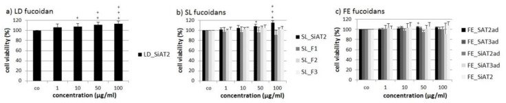
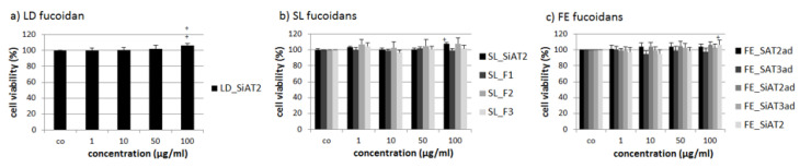
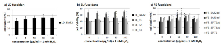
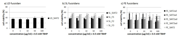
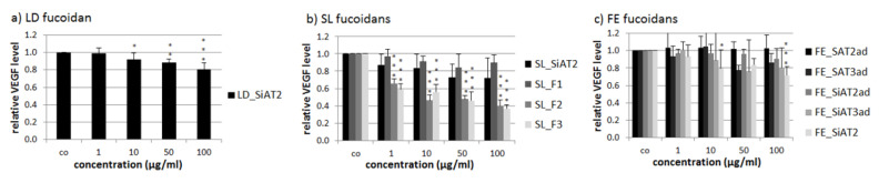
Fucoidans from brown seaweeds are promising substances as potential drugs against age-related macular degeneration (AMD). The heterogeneity of fucoidans requires intensive research in order to find suitable species and extraction methods. Ten different fucoidan samples extracted enzymatically from Laminaria digitata (LD), Saccharina latissima (SL) and Fucus distichus subsp. evanescens (FE) were tested for toxicity, oxidative stress protection and VEGF (vascular endothelial growth factor) inhibition. For this study crude fucoidans were extracted from seaweeds using different enzymes and SL fucoidans were further separated into three fractions (SL_F1-F3) by ion-exchange chromatography (IEX). Fucoidan composition was analyzed by high performance anion exchange chromatography (HPAEC) after acid hydrolysis. The crude extracts contained alginate, while two of the fractionated SL fucoidans SL_F2 and SL_F3 were highly pure. Cell viability was assessed with an 3-(4,5-dimethylthiazol-2-yl)-5-(3-carboxymethoxyphenyl)-2-(4-sulfophenyl)-2H-tetrazolium (MTS) assay in OMM-1 and ARPE-19. Protective effects were investigated after 24 h of stress insult in OMM-1 and ARPE-19. Secreted VEGF was analyzed via ELISA (enzyme-linked immunosorbent assay) in ARPE-19 cells. Fucoidans showed no toxic effects. In OMM-1 SL_F2 and several FE fucoidans were protective. LD_SiAT2 (Cellic®CTec2 + Sigma-Aldrich alginate lyase), FE_SiAT3 (Cellic® CTec3 + Sigma-Aldrich alginate lyase), SL_F2 and SL_F3 inhibited VEGF with the latter two as the most effective. We could show that enzyme treated fucoidans in general and the fractionated SL fucoidans SL_F2 and SL_F3 are very promising for beneficial AMD relevant biological activities.

Keywords: Fucus distichus subsp. evanescens; Laminaria digitata; Saccharina latissima; VEGF; age-related macular degeneration; enzymatic purification; fucoidan; fucose; oxidative stress; retinal pigment epithelium.


Conflict of interest statement

The authors declare no conflict of interest.

Figures

Figure 1
Figure 1
The cell viability of the uveal melanoma cell line OMM-1 was assessed after treatment for 24 h with Laminaria digitata (LD) fucoidan (a), Saccharina latissima (SL) fucoidans (b) and Fucus distichus subsp. evanescens (FE) fucoidans (c) extracted with SiAT2/3 or SAT2/3 (SiAT2/3 = Cellic®CTec2 or 3 enzyme mix + Sigma-Aldrich alginate lyase (SigmALy), SAT2/3 = Cellic®CTec2 or 3 enzyme mix + alginate lyase expressed from Sphingomonas sp. (SALy), ad = acid treatment and dialysis). Also, three SL ion-exchange chromatography (IEX) fractions (SL_F1, SL_F2 and SL_F3) were invastigated. Cell viability was analyzed with a MTS (3-(4,5-Dimethylthiazol-2-yl)-5-(3-carboxymethoxyphenyl)-2-(4-sulfophenyl)-2H-tetrazolium) assay and is shown as the mean and standard deviation in relation to the 100% control. Significance was determined with ANOVA; + p < 0.05, ++ p < 0.01, +++ p < 0.001 compared to control (n ≥ 4; number of independent experiments). No fucoidan exhibited antiproliferative effects.
Figure 2
Figure 2
The cell viability of the human RPE cell line ARPE-19 was assessed after treatment for 24 h with LD fucoidan (a), SL fucoidans (b) and FE fucoidans (c). Cell viability was analyzed with a MTS assay and is shown as the mean and standard deviation in relation to the 100% control. Significance was determined with ANOVA; + p < 0.05, ++ p < 0.01 compared to control (n ≥ 4, number of independent experiments). No fucoidan showed antiproliferative effects.
Figure 3
Figure 3
OMM-1 cell survival after 30 min treatment with LD fucoidan (a), SL fucoidans (b) and FE fucoidans (c) and 24 h stress insult with 1 mM H2O2, which reduced cell viability to at least 60% in all cases. Viability was determined with MTS assay. Values are pictured as the mean and standard deviation in relation to an untreated control (100%). Significance was evaluated via ANOVA; + p < 0.05, ++ p < 0.01, +++ p < 0.001 versus 0 µg/mL fucoidan + 1 mM H2O2 (n ≥ 4, number of independent experiments).
Figure 4
Figure 4
ARPE-19 cell survival after 30 min treatment with LD fucoidan (a), SL fucoidans (b) and FE fucoidans (c) and 24 h stress insult with 0.5 mM TBHP (tert-butyl hydroperoxide), which reduced cell viability below 60% in all cases. Viability was determined with MTS assay. Values are pictured as the mean and standard deviation in relation to an untreated control (100%). Significance was evaluated via ANOVA; + / * p < 0.05, ** p < 0.01, *** p < 0.001 versus 0 µg/mL fucoidan + 0.5 mM TBHP (n ≥ 4, number of independent experiments).
Figure 5
Figure 5
Secreted VEGF (vascular endothelial growth factor) of ARPE-19 after three days of incubation with 1, 10, 50 and 100 µg/mL LD fucoidan (a), SL fucoidans (b) and FE fucoidans (c). VEGF amount was determined with ELISA and normalized to cell survival, making a quotient of VEGF and cell viability. 10–100 µg/mL LD_SiAT2 and 1–100 µg/mL SL_F2 and SL_F3 decreased VEGF significantly. Significant values were analyzed with ANOVA, * p < 0.05, ** p < 0.01, *** p < 0.001 compared to the control (n ≥ 4, number of independent experiments).

References

    1. Wong W.L., Su X., Li X., Cheung C.M.G., Klein R., Cheng C.-Y., Wong T.Y. Global prevalence of age-related macular degeneration and disease burden projection for 2020 and 2040: A systematic review and meta-analysis. Lancet Glob. Health. 2014;2:e106–e116. doi: 10.1016/S2214-109X(13)70145-1. - DOI - PubMed
    1. Miller J.W. Age-related macular degeneration revisited--piecing the puzzle: The LXIX Edward Jackson memorial lecture. Am. J. Ophthalmol. 2013;155:1–35.e13. doi: 10.1016/j.ajo.2012.10.018. - DOI - PubMed
    1. Ding X., Patel M., Chan C.-C. Molecular pathology of age-related macular degeneration. Prog. Retin. Eye Res. 2009;28:1–18. doi: 10.1016/j.preteyeres.2008.10.001. - DOI - PMC - PubMed
    1. Klettner A. Oxidative stress induced cellular signaling in RPE cells. Front. Biosci. 2012;4:392–411. doi: 10.2741/s275. - DOI - PubMed
    1. Hageman G.S., Anderson D.H., Johnson L.V., Hancox L.S., Taiber A.J., Hardisty L.I., Hageman J.L., Stockman H.A., Borchardt J.D., Gehrs K.M., et al. A common haplotype in the complement regulatory gene factor H (HF1/CFH) predisposes individuals to age-related macular degeneration. Proc. Natl. Acad. Sci. USA. 2005;102:7227–7232. doi: 10.1073/pnas.0501536102. - DOI - PMC - PubMed

MeSH terms