Review
. 2020 Apr;55(2):197-218.
doi: 10.1080/10409238.2020.1768207. Epub 2020 May 28.

Retinol-binding protein 2 (RBP2): biology and pathobiology

Affiliations
Review

Retinol-binding protein 2 (RBP2): biology and pathobiology

William S Blaner et al. Crit Rev Biochem Mol Biol. 2020 Apr.

Abstract

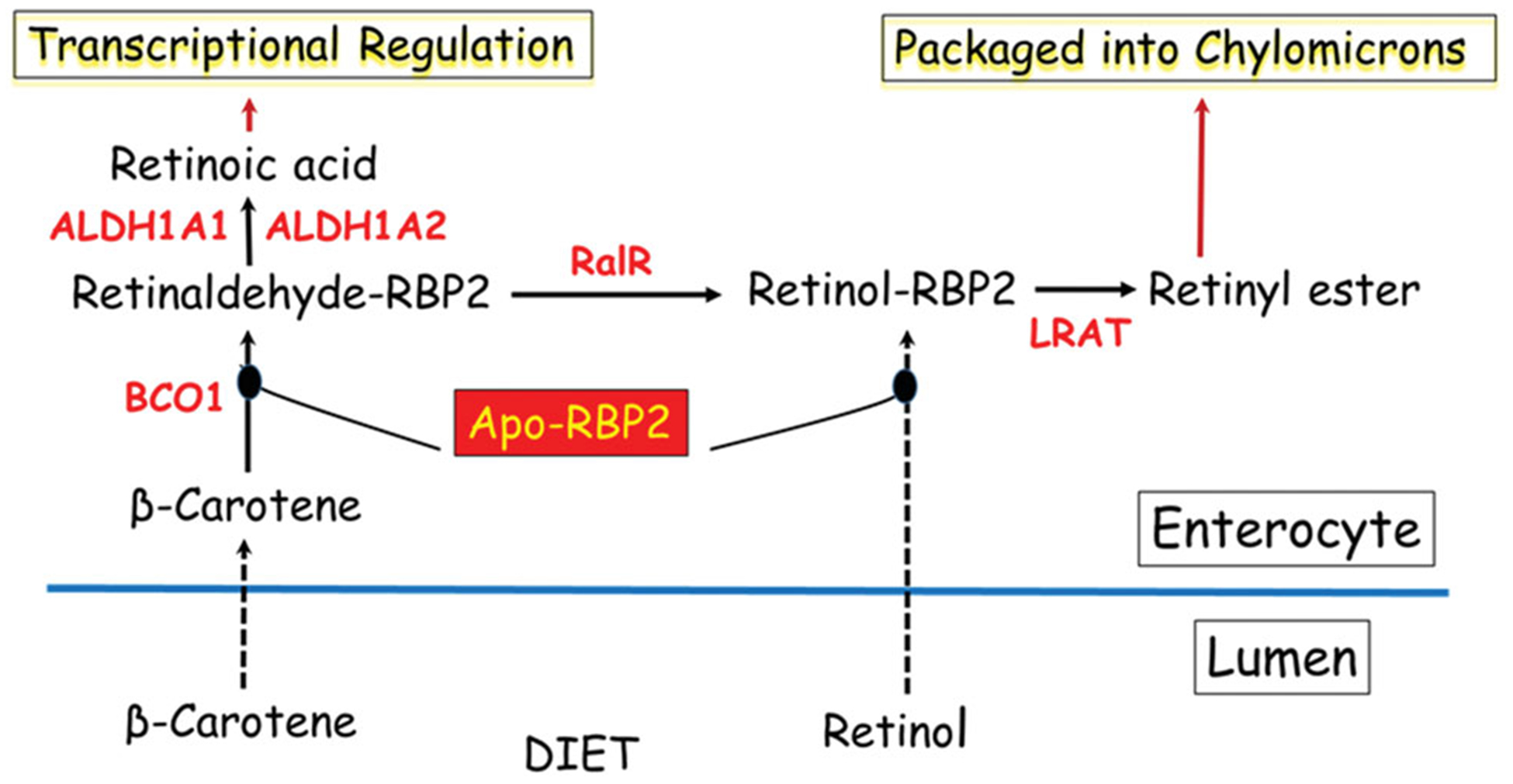
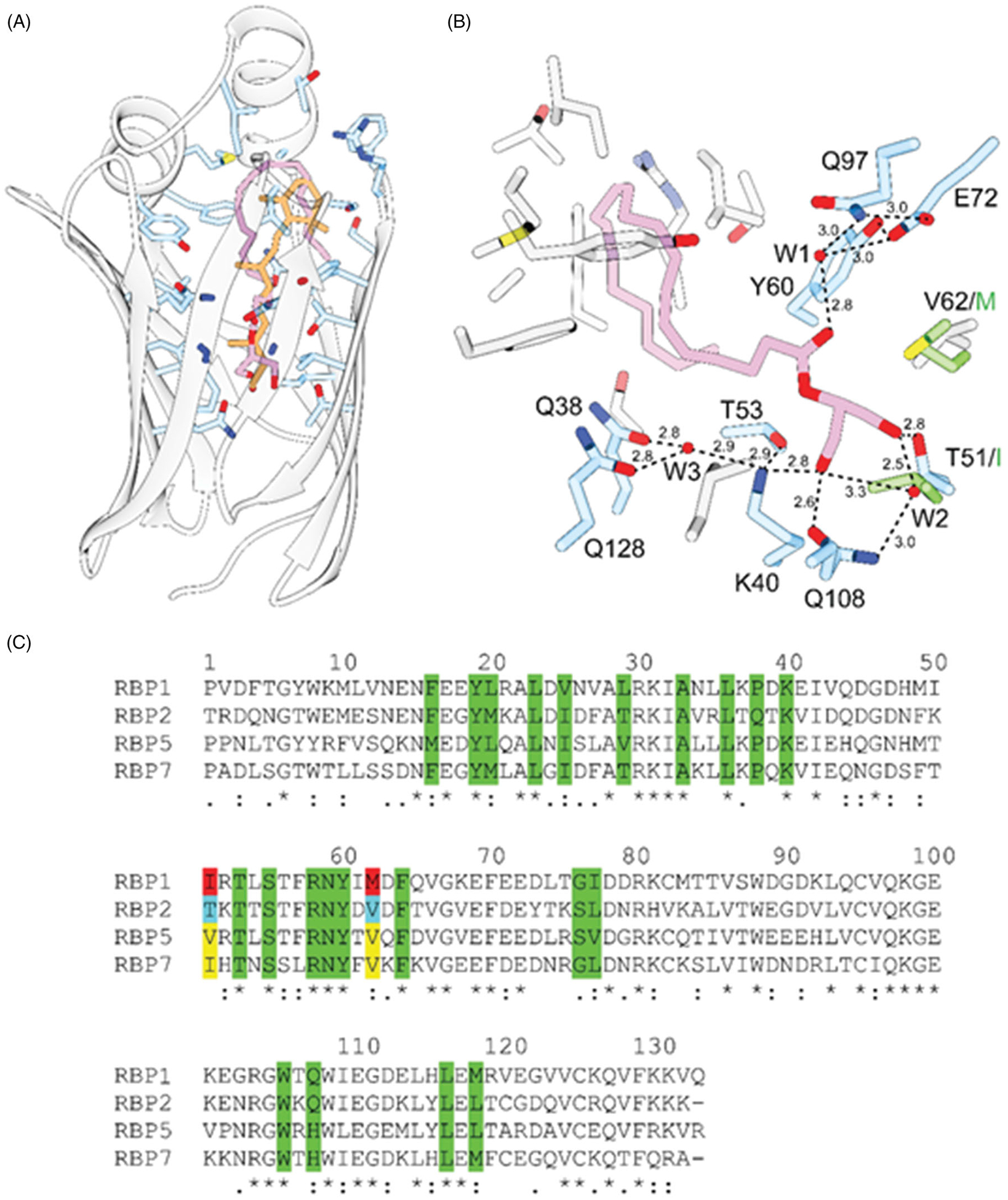
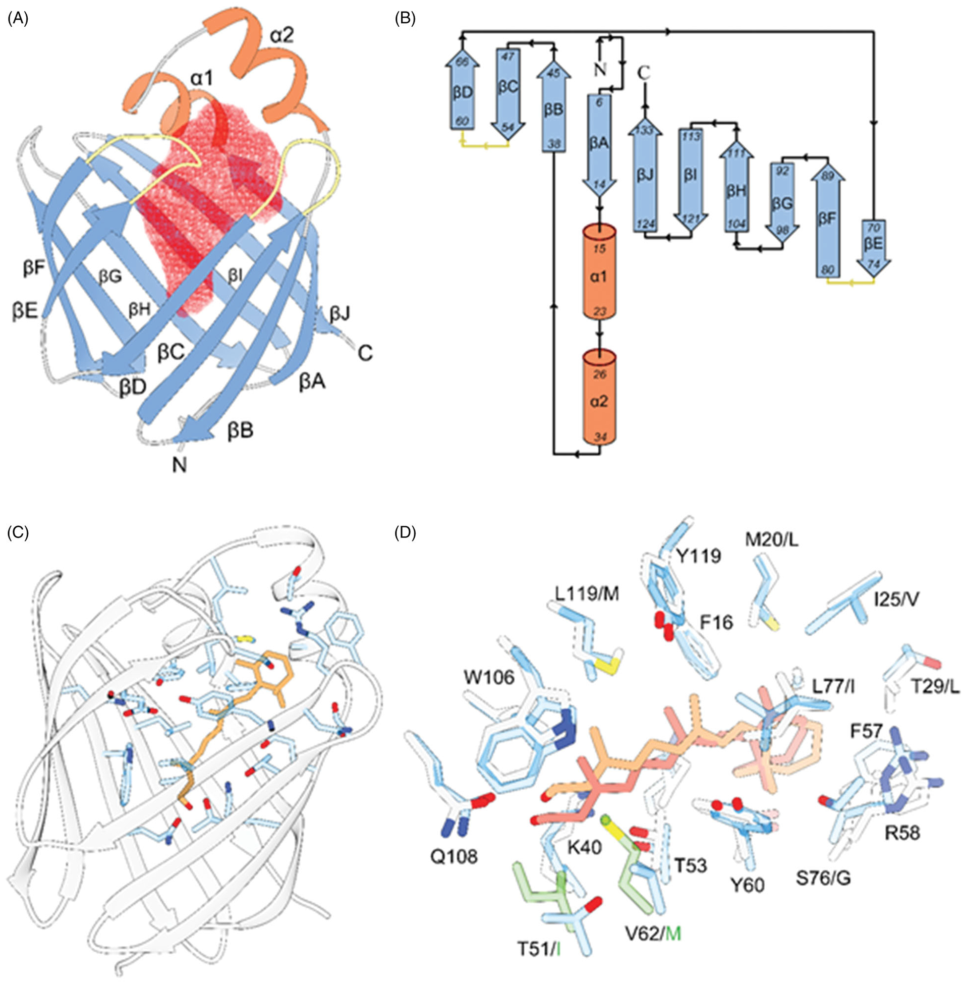
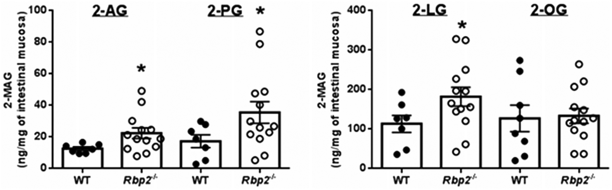
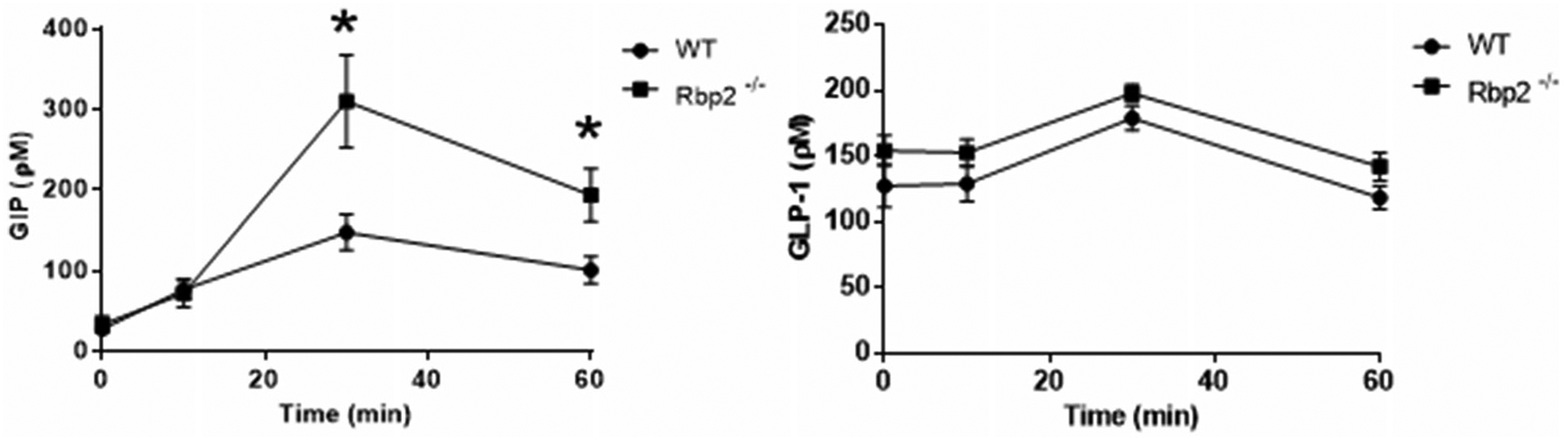
Retinol-binding protein 2 (RBP2; originally cellular retinol-binding protein, type II (CRBPII)) is a 16 kDa cytosolic protein that in the adult is localized predominantly to absorptive cells of the proximal small intestine. It is well established that RBP2 plays a central role in facilitating uptake of dietary retinoid, retinoid metabolism in enterocytes, and retinoid actions locally within the intestine. Studies of mice lacking Rbp2 establish that Rbp2 is not required in times of dietary retinoid-sufficiency. However, in times of dietary retinoid-insufficiency, the complete lack of Rbp2 gives rise to perinatal lethality owing to RBP2 absence in both placental (maternal) and neonatal tissues. Moreover, when maintained on a high-fat diet, Rbp2-knockout mice develop obesity, glucose intolerance and a fatty liver. Unexpectedly, recent investigations have demonstrated that RBP2 binds long-chain 2-monoacylglycerols (2-MAGs), including the canonical endocannabinoid 2-arachidonoylglycerol, with very high affinity, equivalent to that of retinol binding. Crystallographic studies establish that 2-MAGs bind to a site within RBP2 that fully overlaps with the retinol binding site. When challenged orally with fat, mucosal levels of 2-MAGs in Rbp2 null mice are significantly greater than those of matched controls establishing that RBP2 is a physiologically relevant MAG-binding protein. The rise in MAG levels is accompanied by elevations in circulating levels of the hormone glucose-dependent insulinotropic polypeptide (GIP). It is not understood how retinoid and/or MAG binding to RBP2 affects the functions of this protein, nor is it presently understood how these contribute to the metabolic and hormonal phenotypes observed for Rbp2-deficient mice.

Keywords: Vitamin A; endocannabinoid; enteroendocrine signaling; glucose-dependent insulinotropic polypeptide (GIP) and dietary fat; intestine; monoacylglycerol; obesity; retinoid.


Figures

Figure 1.
Figure 1.
Longstanding understanding of the role of RBP2 in retinoid uptake, metabolism and actions within the absorptive cells (enterocytes) of the small intestine. These arise due to the ability of RBP2 to bind retinol or retinaldehyde with high affinity. This allows apo-RBP2 to bind retinol as it is newly absorbed by enterocytes from the intestinal lumen after arriving there as a component of the diet. Alternatively, apo-RBP2 can bind retinaldehyde formed from newly absorbed β-carotene by β-carotene 15,15′-dioxygenase (BCO1) within the enterocyte. Retinaldehyde bound to RBP2 is a substrate for the intestinal retinaldehyde reductase (RalR) allowing for retinaldehyde conversion to retinol, while bound to RBP2. Retinol bound to RBP2 is a substrate for lecithin:retinol acyltransferase (LRAT) producing retinyl esters that are packaged into nascent chylomicrons for uptake into the body. Thus, retinol or retinaldehyde obtained from the diet is metabolically channeled by RBP2 to the chylomicrons for uptake along with other dietary fat into the circulation. Some retinaldehyde bound to RBP2 is converted to retinoic acid by the actions of retinaldehyde dehydrogenases 1 and 2 (ALDH1A1 and ALDH1A2). The retinoic acid can be used locally to regulate retinoic acid-dependent transcription.
Figure 2.
Figure 2.
Interaction of 2-AG with RBP2. A, the overall orientation of 2-AG (purple) within the binding-pocket of human RBP2 (PDB #6BTH) in comparison to all-trans-retinol (orange) (PDB #4QZT). B, hydrogen bond network that contribute to the high affinity of 2-AG. Hydrogen bonds are indicated by dashed lines. Distances are shown in Å. Ordered water molecules (W) are depicted as red sphered. C, sequence alignment of human RBPs. Amino acid that line the inside of the binding cavities are marked in green. Residues in position 51 and 62 that prevent binding of monoacylglycerols to RBP1 are labeled in red. Yellow background highlights residues found in the sequences of RBP5 and RBP7. They may indicate an alternative ligand specificity to RBP1 and RBP2.
Figure 3.
Figure 3.
The overall structural motive and the mode of all-trans-retinol interaction with RBPs. A, ribbon diagram of human RBP2 (PDB #6BTH). The red mesh represents the ligand-binding cavity inside the β-barrel composed of ten antiparallel β-strands. The entry portal region is defined by two α-helices (shown in orange) and loops connecting and β-strands C-D and E-F (colored yellow). B, topology diagrams for the RBPs. The color scheme is identical as in panel A. C, position of the retinoid moiety within the binding pocket of RBP2 (PDB #4QZT). The side chains of amino acids in the vicinity of all-trans-retinol (orange) are highlighted in blue. D, overlay of the binding sites structures and ligand orientation found in human RBP1 (PDB #5HBS) and human RBP2 (PDB #4QZT). The side chains of RBP1 and RBP2 are colored gray and blue, respectively. The position of all-trans-retinol seen in RBP1 is shown in light orange, whereas the retinoid moiety found in RBP2 is colored in pink. The side chains shown in green correspond to the substitutions of T51/I and V62/M in RBP1 that contribute to the different in the position of the ligand.
Figure 4.
Figure 4.
An oral fat challenge administered to Rbp2−/− mice results in elevated 2-MAG levels in intestinal absorptive cells. Two hours after administration of an oral challenge with corn oral, levels of 2-arachidonoylglycerol (2-AG) 2-palmitoylglycerol (2-PG), and 2-lineoylglycerol (2-LG) are significantly elevated in enterocyte scrapings for Rbp2−/− mice compared to matched wild type (WT) mice. Enterocyte levels of 2-oleoylglycerol (2-OG) were not significantly different for the two groups. *p < 0.05. (Taken from Lee et al. 2020).
Figure 5.
Figure 5.
Time course showing plasma levels of glucose-dependent insulinotropic polypeptide (GIP) (left) and glucagon-like protein-1 (GLP-1) (right) following an oral challenge with corn oil. Plasma GIP levels are significantly elevated 30 and 60 min after the challenge for chow fed 6- to 7-month-old Rbp2−/− mice compared with matched wild-type mice. No statistically significant differences in plasma GLP-1 levels were observed for the same mice. *p < 0.05.

References

    1. Agace WW, Persson EK. 2012. How vitamin A metabolizing dendritic cells are generated in the gut mucosa. Trends Immunol. 33(1):42–48. - PubMed
    1. Bashor MM, Toft DO, Chytil F. 1973. In vitro binding of retinol to rat-tissue components. Proc Natl Acad Sci USA. 70(12):3483–3487. - PMC - PubMed
    1. Bhat PV. 1998. Retinal dehydrogenase gene expression in stomach and small intestine of rats during postnatal development and in vitamin A deficiency. FEBS Lett. 426(2):260–262. - PubMed
    1. Blaner WS, Li Y. 2015. Vitamin A metabolism, storage and tissue delivery mechanisms In: Doll e P, Niederreither K, editors. The retinoids; biology, biochemistry, and disease. Hoboken (NJ): Wiley Blackwell; p. 3–34.
    1. Calanna S, Christensen M, Holst JJ, Laferrère B, Gluud LL, Vilsbøll T, Knop FK. 2013. Secretion of glucose-dependent insulinotropic polypeptide in patients with type 2 diabetes: systematic review and meta-analysis of clinical studies. Diabetes Care 36(10):3346–3352. - PMC - PubMed

Publication types

MeSH terms