. 2020 Jun 16;117(24):13468-13479.
doi: 10.1073/pnas.1908232117. Epub 2020 May 28.

PI(4,5)P2-dependent regulation of exocytosis by amisyn, the vertebrate-specific competitor of synaptobrevin 2

Affiliations

PI(4,5)P2-dependent regulation of exocytosis by amisyn, the vertebrate-specific competitor of synaptobrevin 2

Ilona Kondratiuk et al. Proc Natl Acad Sci U S A. .

Abstract

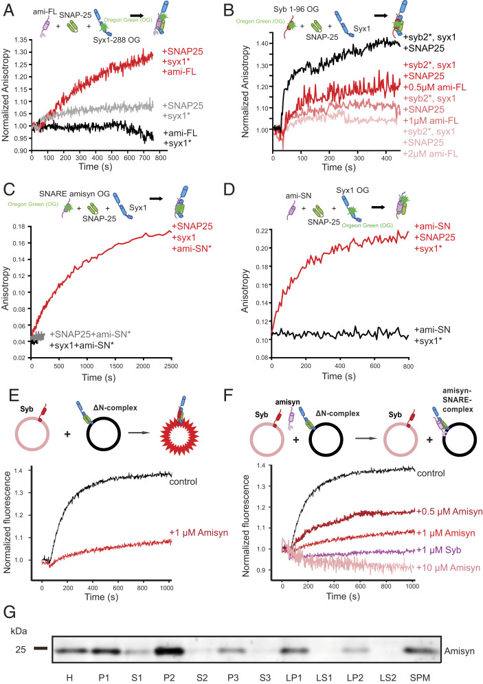
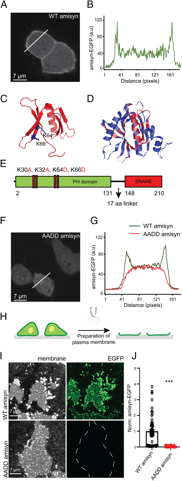
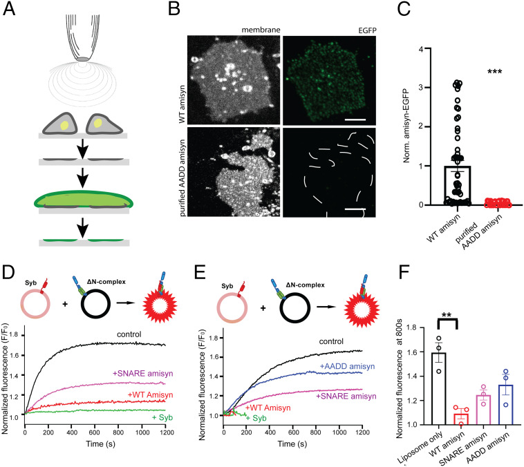
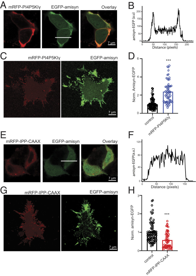
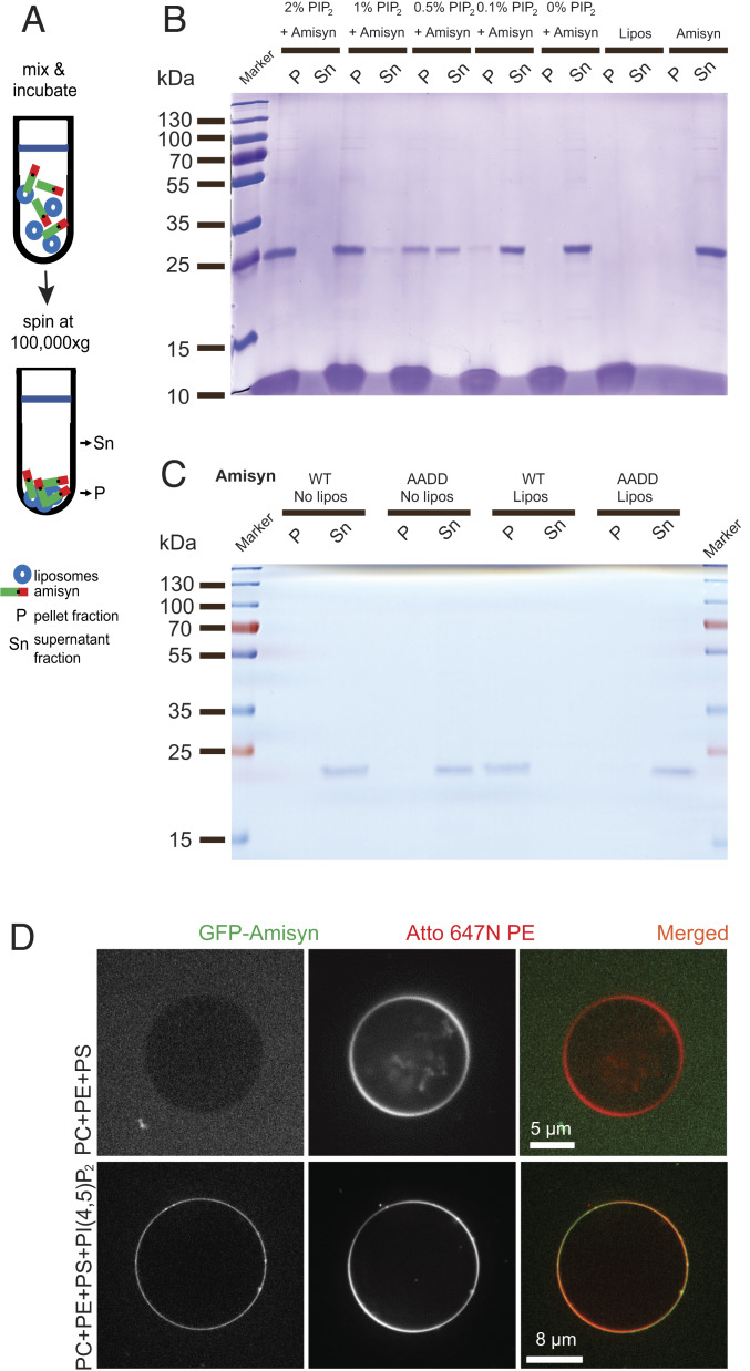
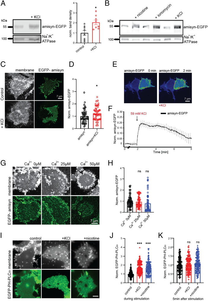
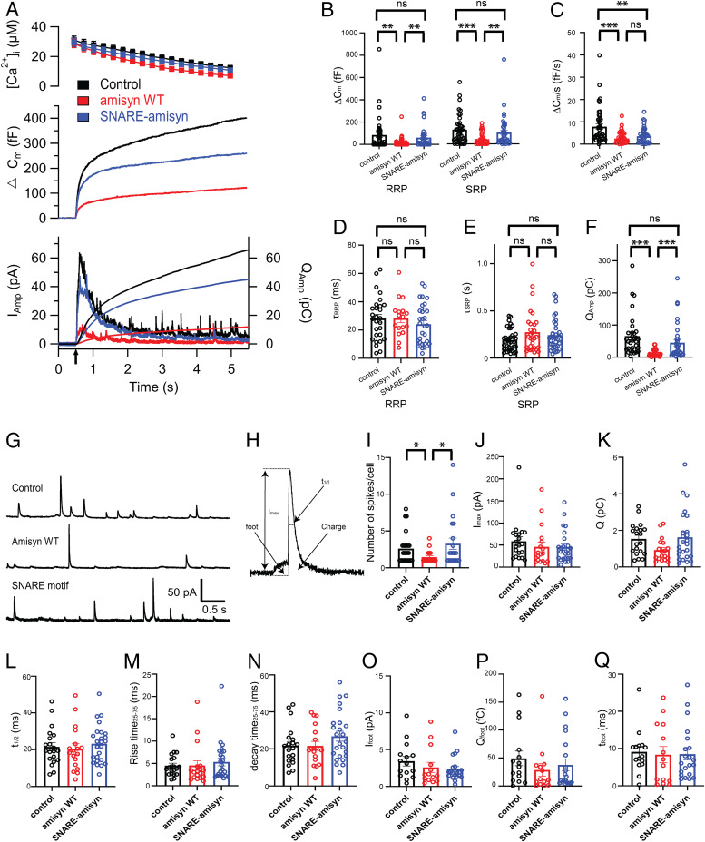
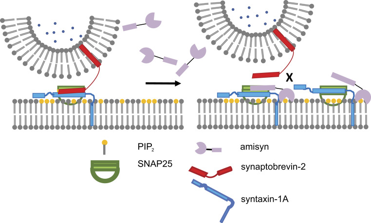
The functions of nervous and neuroendocrine systems rely on fast and tightly regulated release of neurotransmitters stored in secretory vesicles through SNARE-mediated exocytosis. Few proteins, including tomosyn (STXBP5) and amisyn (STXBP6), were proposed to negatively regulate exocytosis. Little is known about amisyn, a 24-kDa brain-enriched protein with a SNARE motif. We report here that full-length amisyn forms a stable SNARE complex with syntaxin-1 and SNAP-25 through its C-terminal SNARE motif and competes with synaptobrevin-2/VAMP2 for the SNARE-complex assembly. Furthermore, amisyn contains an N-terminal pleckstrin homology domain that mediates its transient association with the plasma membrane of neurosecretory cells by binding to phospholipid PI(4,5)P2 However, unlike synaptrobrevin-2, the SNARE motif of amisyn is not sufficient to account for the role of amisyn in exocytosis: Both the pleckstrin homology domain and the SNARE motif are needed for its inhibitory function. Mechanistically, amisyn interferes with the priming of secretory vesicles and the sizes of releasable vesicle pools, but not vesicle fusion properties. Our biochemical and functional analyses of this vertebrate-specific protein unveil key aspects of negative regulation of exocytosis.

Keywords: PI(4,5)P2; SNARE complex; autism spectrum disorders; exocytosis inhibition; tomosyn.


Conflict of interest statement

The authors declare no competing interest.

Figures

Fig. 1.
Fig. 1.
Amisyn forms SNARE complex with syntaxin-1 and SNAP-25 and inhibits liposome fusion in vitro. (A) Fluorescence anisotropy of syntaxin-1 (syx11−288OG; 1 μM) revealed interaction with full-length amisyn (ami-FL; 1 μM) in the presence of SNAP-25 (1.5 μM) (red), versus SNAP-25 (gray), and amisyn alone (black trace). (B) Fluorescence anisotropy of synaptobrevin-2 (syn21−96OG) revealed interaction with syntaxin-1 in the presence of SNAP-25 (1.5 μM) and full-length amisyn (0.5, 1, or 2 μM). Competition between amisyn and synaptobrevin-2 is demonstrated by decrease in anisotropy upon adding amisyn in a concentration-dependent manner. (C) Anisotropy of the SNARE motif of amisyn (100 nM), labeled at cysteine-210 with Oregon green-488, did not change by addition of the H3-syntaxin-1a (syx1, 1 μM) or SNAP-25 (SNAP25; 1.5 μM). In contrast, increased anisotropy was observed when both syx1 and SNAP-25 were added. (D) Anisotropy of H3-syntaxin-1a (syx1), labeled at position 197 with Oregon green-488 (syx1197OG), did not change by addition of the SNARE motif of amisyn (Ami-SN). Increased anisotropy was evident when both ami-SN and SNAP-25 (SNAP25) were added to syx1197OG. (E) In the presence of amisyn, liposome fusion is severely impaired. Schematic representation of the liposome fusion assay is shown above the graph. Donor liposomes contain Syb-2 (1–116) and two fluorophores, nitrobenzoxadiazole (NBD) and Rhodamine, coupled to lipids, thereby quenching their fluorescence. Acceptor liposomes contain ∆N ternary complexes of SNAP-25, syntaxin-1a, and a C-terminal fragment of Syb-2. When donor (200 nM) and acceptor (200 nM) liposomes are mixed, they fuse due to trans-SNARE complex formation, which allows de-quenching of the NBD fluorescence and kinetic and quantitative measurements of the fusion process. Amisyn caused less dequenching, indicative of inhibition of the fusion process. (F) Inhibition of liposome fusion by amisyn is concentration-dependent. Monitoring of NBD-dequenching fluorescence after mixing liposomes revealed that higher concentrations of amisyn blocked liposome fusion more efficiently. Soluble synaptobrevin-2, which competes with synaptobrevin-2 on the liposomes for the SNARE complex formation, was added as a control. (G) Fractionation of mouse brain to define the subcellular distribution of amisyn. See also SI Appendix, Fig. S1F. Abbreviations: H, mouse brain homogenate; P1, 1,400 × g pellet; S1, supernatant, further centrifuged (13,800 × g, 10 min) to yield pellet P2: crude synaptosomes. P2 was lysed and centrifuged (32,800 × g, 20 min) to yield pellet LP1: crude synaptic plasma membranes and supernatant LS2: synaptic vesicles and synaptosomal cytosol. S2: crude cytosol further centrifuged (165,000 × g, 1 h) to obtain P3: membranes and S3: cytosol. Fraction LS1 was centrifuged (165,000 × g, 1 h) to yield pellet containing crude synaptic vesicles (LP2) and supernatant containing synaptosomal cytosol (LS2). SPM contains isolated synaptic membranes after sucrose gradient centrifugation. Amisyn was predominantly found in membrane fractions.
Fig. 2.
Fig. 2.
Amisyn binds to membranes by its N-terminal PH-domain. (A) Amisyn-EGFP distribution in PC12 cells revealing amisyn enrichment at the plasma membrane. Cells were fixed and imaged 20 h posttransfection. An identical result was observed in six independent experiments. (B) The fluorescence intensity line profile according to the white line in A. (C) Model of the N-terminal domain of amisyn that assembles into a PH-like domain. The essential Lysine residues (K64 and K66) are marked in blue. (D) Alignment of the identified PH domain of amisyn (red) with that of PLCɗ1 (blue) as template. (E) Schematic representation of amisyn with the PH and SNARE domains separated by the 17-amino acid linker sequence. The mutant AADD-amisyn protein contained four point-mutations in the PH domain: K30A, K32A, K64D, K66D. (F) The mutant AADD-amisyn failed to bind to the plasma membrane. Representative confocal image of AADD amisyn-EGFP expressed in PC12 cells. Cells were fixed and imaged 20 h posttransfection. The experiment was repeated three times. (G) The fluorescence intensity profiles of PC12 cells expressing WT amisyn-EGFP (green trace) and AADD amisyn-EGFP (red trace; from the white line in F). (H) Schematic of isolated PC12 plasma membrane sheet preparation by sonication. (I) Isolated membrane sheets from PC12 cells expressing either WT or AADD amisyn-EGFP (indicated by the captions on the left). Representative images are shown of the membranes (Left) and the associated fluorescence (Right). (J) Quantification of the fluorescence signals (as shown in I) of WT and AADD amisyn-EGFP. Three independent experiments, WT (31 cells) and AADD (33 cells). Unpaired two-sided t test, mean ± SEM, ***P < 0.001.
Fig. 3.
Fig. 3.
Recombinant amisyn binds to plasma membranes in vitro. (A) Schematic of PC12 plasma membrane sheet preparation by sonication and subsequent immediate incubation with purified recombinant amisyn protein. (B) Binding of recombinant WT and AADD-amisyn to membranes from PC12 cells. The purified WT amisyn-EGFP (1 μM) or AADD amisyn-EGFP (1 μM) were incubated with freshly prepared membranes at room temperature. Images show WT but not AADD-amisyn-EGFP bound to the membranes (visualized by trimethylamine-diphenylhexatriene [TMA-DPH]). (Scale bar, 5 μm.) (C) Quantitative analysis of data as shown in B. Unpaired two-sided t test, At least 49 sheets/conditions from three experiments; mean ± SEM, ***P < 0.001. (D) Representative experiment showing full-length WT amisyn inhibiting liposome fusion more efficiently than its SNARE domain. Fluorescence dequenching of NBD fluorophore-labeled liposomes containing synaptobrevin-2 (Syb) after mixing with liposomes containing the ΔN complex. Soluble synaptobrevin-2 that competes with synaptobrevin-2 on the liposomes for the SNARE complex formation was added as a control. (E) Representative experiment showing that WT amisyn inhibits liposome fusion more efficiently than AADD amisyn mutant. Fluorescence dequenching of NBD fluorophore-labeled liposomes containing synaptobrevin-2 (Syb) after mixing with liposomes containing the ΔN complex. (F) Fluorescence dequenching of NBD fluorophore labeled liposomes at 800s reveals that WT amisyn inhibits liposome fusion more efficiently than its SNARE domain alone or AADD-amisyn. Three independent experiments; mean ± SEM.
Fig. 4.
Fig. 4.
PI(4,5)P2 levels control membrane binding of amisyn in PC12 cells. (A) Representative confocal images of PC12 cells expressing either amisyn-EGFP or mRFP-PI4P5KIγ, or both to increase PI(4,5)P2 in inner leaflet of membrane, resulting in more amisyn associated with the plasma membrane. (B) Fluorescence profile of amisyn-EGFP expressing cell through a white line in A. (C) Plasma membrane sheets isolated from cotransfected PC12 cells as in A contain more amisyn-EGFP. (D) Quantitation of amisyn-EGFP fluorescence on the plasma membrane sheets isolated from transfected PC12 cells. At least 50 sheets/conditions, two independent experiments; mean ± SEM, unpaired two-sided t test, ***P < 0.001. (E) Confocal images of PC12 cells expressing either amisyn-EGFP or mRFP-IPP-CAAX or both, to decrease levels of PI(4,5)P2 in the inner leaflet of the plasma membranes, resulting in less amisyn-EGFP bound to the plasma membrane. (F) Fluorescence profile through an amisyn-EGFP–expressing cell through a white line in E. (G) Membrane sheets isolated from cotransfected PC12 cells as in E contain less amisyn-EGFP. (H) Quantitative determination of amisyn-EGFP fluorescence intensity on membrane sheets from transfected PC12 cells. At least 50 sheets/conditions, three experiments. Mean ± SEM, unpaired two-sided t test, ***P < 0.001.
Fig. 5.
Fig. 5.
Liposome binding of amisyn is dependent on PI(4,5)P2. (A) Schematic representation of a cosedimentation assay. (B) Representative cosedimentation of amisyn with liposomes depends on their PI(4,5)P2 content (shown in captions over each lane). Representative SDS/PAGE gel (12%) shows that PI(4,5)P2 levels in liposomes correlate with more amisyn bound to liposomes cosedimenting in the pellets (P) relative to the supernatant (Sn). Three independent experiments were performed. (C) Representative cosedimentation assay shows that AADD amisyn mutant does not bind well to PI(4,5)P2-containing liposomes (2%). Representative SDS/PAGE gel (12%) of sedimentation assay. P, pellet; Sn, supernatant; three independent experiments were performed. (D) Recombinant amisyn-EGFP bound only to liposomes containing PI(4,5)P2. Representative confocal images of liposomes without or containing PI(4,5)P2 (captions on the left). Two experiments with independently purified amisyn-EGFP were performed, each time with several technical replicates.
Fig. 6.
Fig. 6.
Stimulation recruits amisyn-EGFP to the plasma membrane of PC12 cells. (A) Membranes isolated from PC12 cells after stimulation (59 mM KCl, 5 s) contain more amisyn-EGFP, relative to membranes isolated from naive cells. Na+/K+ ATPase is used as internal loading control and membrane marker. (Left) Representative Western blots from eight experiments. (Right) Quantification. Mean ± SEM, unpaired two-sided t test, *P < 0.1. (B) Representative Western blot of membranes isolated from naive PC12 cells and after stimulation as shown in the captions (100 μM nicotine, 1 μM ionomycin, 59 mM KCl, 5 s). Na+/K+ ATPase was used as the internal loading control and membrane marker. (C) Membrane sheets generated from stimulated PC12 cells (59 mM KCl, 5 s) contain more amisyn-EGFP than membrane sheets from naive cells. (D) Fluorescence quantified from samples as in C (n = 41 sheets/conditions, 3 experiments). Mean ± SEM; unpaired two-sided t test, *P < 0.1. (E) Representative confocal images of PC12 cells before and after stimulation (59 mM KCl) demonstrating that stimulation recruited amisyn to plasma membranes. (F) Time-course of amisyn-EGFP fluorescence on the plasma membrane in living PC12 cells stimulated with 59 mM KCl. Mean of 15 cells from 3 experiments ± SEM (G) Plasma membrane sheets from naive PC12 cells were incubated with recombinant amisyn-EGFP (3 μM) and different calcium ion concentrations (captions). (Upper) TMA-DPH–stained isolated plasma membranes; (Lower) isolated plasma membranes with bound recombinant amisyn-EGFP. (H) Quantification of amisyn fluorescence (as on samples in G) revealed that recruitment of amisyn-EGFP to the plasma membrane was not mediated by calcium ions. At least 46 sheets/condition from 3 experiments. Mean ± SEM; one-way ANOVA with Tukey’s post hoc test; ns, not significant. (I) Plasma membrane sheets isolated from stimulated PC12 cells (59 mM KCl, 5 s) were incubated with recombinant EGFP-PH-PLCɗ1 (3 μM) for 60 s, washed and fixed. (Upper) TMA-DPH dye stains isolated plasma membranes; (Lower) isolated plasma membranes with bound recombinant EGFP-PH-PLCɗ1. (Scale bar, 5 µm.) (J) Quantification of EGFP-PH-PLCɗ1 fluorescence (as on samples in I) revealed elevated PI(4,5)P2 levels in the plasma membranes of stimulated cells. At least 144 sheets/conditions from 3 experiments. Mean ± SEM; one-way ANOVA with Tukey’s post hoc test, ***P < 0.001. (K) Plasma membrane sheets were isolated from PC12 cells 5 min poststimulation (59 mM KCl), immediately incubated with recombinant EGFP-PH-PLCɗ1 (3 μM) for 60 s, washed and fixed. Quantification of EGFP-PH-PLCɗ1 fluorescence revealed no change in the levels of PI(4,5)P2 in the plasma membranes between nonstimulated and stimulated cells. At least 144 sheets/conditions from 3 experiments. Mean ± SEM; one-way ANOVA with Tukey’s post hoc test; ns, not significant.
Fig. 7.
Fig. 7.
Amisyn, but not amisyn-SNARE domain, reduced number of released vesicles but did not alter rates of vesicle fusion in bovine chromaffin cells. (AF) Exocytosis induced by UV-flash photolysis of caged calcium ions (stimulus #1, arrow) was reduced in amisyn-loaded chromaffin cells (red trace) compared to control cells (black trace). Cells loaded with amisyn-SNARE protein (blue trace) did not differ significantly from control cells (with an exception of sustained release): 42 control cells, (black); 38 amisyn-loaded cells (red); 38 amisyn-SNARE–loaded cells (blue) from 5 independent experiments. Kruskal–Wallis test with Dunn’s multiple comparison test; ns, non significant; **P < 0.01, ***P < 0.001. (A, Top) Intracellular calcium level increase induced by flash photolysis (at t = 0.5 s). (Middle) Averaged traces of membrane capacitance changes upon Ca2+-induced exocytosis. (Bottom) Mean amperometric current (Iamp; left axis) and cumulative charge (right axis). (B and C) Exponential fitting of the capacitance traces revealed changes in RRP and SRP size and sustained phase of release (C). Note the marked reduction in exocytosed vesicles in amisyn-loaded cells. (D and E) Fusion kinetics of vesicles from RRP and SRP pools were not altered. (F) Reduced detection of catecholamines in chromaffin cells loaded with amisyn by amperometry: Cumulative charge during 5 s after stimulation. (GQ) Single-spike amperometry analysis revealed problems in vesicle fusion, but no alterations in the stability of the fusion pore. (G) Exemplary traces from single-spike amperometric recordings of control, amisyn WT, and amisyn-SNARE domain-injected adrenal chromaffin cells. (H) Schematic of analyzed amperometric spike parameters. (I) Number of fusion events per cell was significantly reduced in chromaffin cells injected with full-length amisyn. Each spot in the dot plot represents a mean from an analyzed cell. Four experiments and four independent cell preparations, total of control (20 cells), amisyn WT (18 cells), and amisyn-SNARE domain (25 cells). Mean ± SEM. One-way ANOVA with Tukey’s post hoc test, *P < 0.05. (JN) Single-spike amplitude (J), charge (K), and the kinetics of single fusion events, such as duration at half-maximal amplitude (L), rise time (M), and decay time (N), were unchanged. For number of cells and statistics, see I. (OQ) The stability of the fusion pore was not altered, as revealed by unchanged foot amplitude (O), foot charge (P), and foot duration (Q). Four experiments and four independent cell preparations, total of control (15 cells), amisyn WT (14 cells), and amisyn-SNARE domain (21 cells). Mean ± SEM. One-way ANOVA with Tukey’s post hoc test, *P < 0.05.
Fig. 8.
Fig. 8.
Model of role of amisyn in secretory vesicle exocytosis. Amisyn acts as a negative regulator of SNARE-complex assembly by competing with the fusion-active synaptobrevin-2. Formation of the SNARE complex drives membrane fusion, whereby PI(4,5)P2 recruits amisyn to the plasma membrane to compete with synaptobrevin-2 in formation of the SNARE complex and vesicle exocytosis. Since amisyn does not contain a transmembrane domain, it forms a fusion-inactive SNARE complex.

Similar articles

Cited by

References

    1. Terrian D. M., White M. K., Phylogenetic analysis of membrane trafficking proteins: A family reunion and secondary structure predictions. Eur. J. Cell Biol. 73, 198–204 (1997). - PubMed
    1. Weimbs T., Mostov K., Low S. H., Hofmann K., A model for structural similarity between different SNARE complexes based on sequence relationships. Trends Cell Biol. 8, 260–262 (1998). - PubMed
    1. Sutton R. B., Fasshauer D., Jahn R., Brunger A. T., Crystal structure of a SNARE complex involved in synaptic exocytosis at 2.4 A resolution. Nature 395, 347–353 (1998). - PubMed
    1. Fasshauer D., Eliason W. K., Brünger A. T., Jahn R., Identification of a minimal core of the synaptic SNARE complex sufficient for reversible assembly and disassembly. Biochemistry 37, 10354–10362 (1998). - PubMed
    1. Söllner T., Bennett M. K., Whiteheart S. W., Scheller R. H., Rothman J. E., A protein assembly-disassembly pathway in vitro that may correspond to sequential steps of synaptic vesicle docking, activation, and fusion. Cell 75, 409–418 (1993). - PubMed

Publication types

MeSH terms