. 2020 Jul;41(7):1179-1183.
doi: 10.3174/ajnr.A6610. Epub 2020 May 28.

Brain Imaging Use and Findings in COVID-19: A Single Academic Center Experience in the Epicenter of Disease in the United States

Affiliations

Brain Imaging Use and Findings in COVID-19: A Single Academic Center Experience in the Epicenter of Disease in the United States

A Radmanesh et al. AJNR Am J Neuroradiol. 2020 Jul.

Abstract

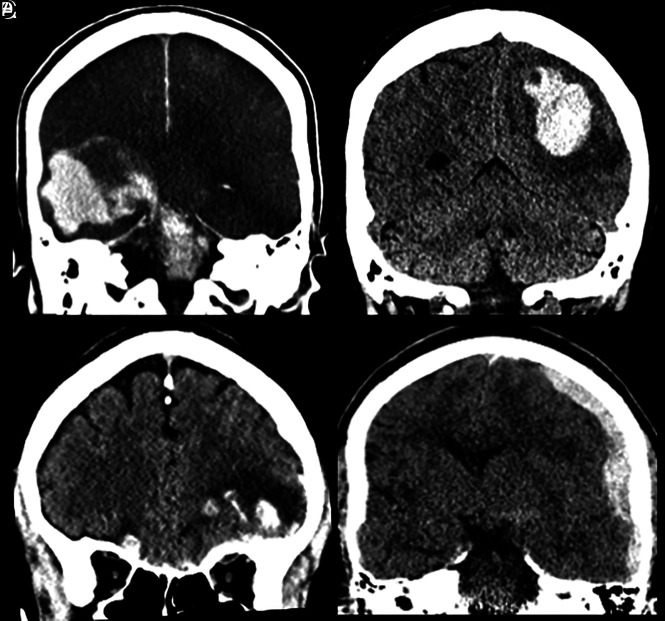
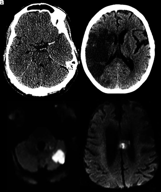
Coronavirus disease 2019 (COVID-19) is a serious public health crisis and can have neurologic manifestations. This is a retrospective observational case series performed March 1-31, 2020, at New York University Langone Medical Center campuses. Clinical and imaging data were extracted, reviewed, and analyzed. Two hundred forty-two patients with COVID-19 underwent CT or MRI of the brain within 2 weeks after the positive result of viral testing (mean age, 68.7 ± 16.5 years; 150 men/92 women [62.0%/38.0%]). The 3 most common indications for imaging were altered mental status (42.1%), syncope/fall (32.6%), and focal neurologic deficit (12.4%). The most common imaging findings were nonspecific white matter microangiopathy (134/55.4%), chronic infarct (47/19.4%), acute or subacute ischemic infarct (13/5.4%), and acute hemorrhage (11/4.5%). No patients imaged for altered mental status demonstrated acute ischemic infarct or acute hemorrhage. White matter microangiopathy was associated with higher 2-week mortality (P < .001). Our data suggest that in the absence of a focal neurologic deficit, brain imaging in patients with early COVID-19 with altered mental status may not be revealing.


Figures

FIG 1.
FIG 1.
Acute intracranial hemorrhage in patients with COVID-19. A, A 74-year-old man with COVID-19, intubated for hypoxic respiratory failure and on heparin due to a history of stented carotid stenosis. On day 10 of intensive care unit admission, he suddenly became unresponsive, and neurologic examination revealed absent brain stem reflexes. Brain CT showed extensive supra- and infratentorial acute hemorrhage with subarachnoid and intraventricular extensions, along with cerebral swelling and hypodensity (likely hypoperfusion injury), as well as uncal, subfalcine, and transtentorial herniations. B, A 61-year-old woman with COVID-19 and compensated hepatic cirrhosis (due to primary sclerosing cholangitis). On day 7 of intensive care unit admission, the patient developed right-sided weakness and numbness. Brain CT showed left parietal intraparenchymal hemorrhage with surrounding vasogenic edema. C, A 68-year-old man with COVID-19 was found fallen. Head CT showed bilateral inferior frontal lobe hemorrhagic contusions and a small subarachnoid hemorrhage, suggesting traumatic brain injury, likely related to the fall. D, A 61-year-old man with COVID-19 who presented after a fall. Brain CT revealed acute left cerebral convexity subdural and left ambient cistern subarachnoid hemorrhage, possibly related to the fall.
FIG 2.
FIG 2.
Acute or subacute infarct in patients with COVID-19. A, A 62-year-old man with COVID-19, intubated for acute hypoxic respiratory failure, who initially presented with left MCA syndrome. Noncontrast head CT showed a dense left MCA sign suggesting a left MCA occlusion, later confirmed on CT angiography and catheter angiography (not shown here). B, A 77-year-old woman with COVID-19 presented with left-sided weakness. Noncontrast CT showed an acute/subacute right MCA territory infarct. C, A 63-year-old COVID-19 patient with ataxia. Brain MRI revealed a patchy acute infarct in the left cerebellar hemisphere. D, A 78-year-old man with COVID-19 presented following an unwitnessed fall. Brain MRI showed left more than right cingulate gyrus and callosal body acute infarct.

Similar articles

Cited by

References

    1. Zhou P, Yang XL, Wang XG, et al. . A pneumonia outbreak associated with a new coronavirus of probable bat origin. Nature 2020;579:270–73 10.1038/s41586-020-2012-7 - DOI - PMC - PubMed
    1. COVID-19 United States Cases by County. Johns Hopkins University of Medicine; May 10, 2020. https://coronavirus.jhu.edu/us-map. Accessed April 25, 2020
    1. COVID-19: Data. NYC Health; City of New York: April 24, 2020. https://www1.nyc.gov/site/doh/covid/covid-19-data.page. Accessed April 25, 2020
    1. Mao L, Jin H, Wang M, et al. . Neurologic manifestations of hospitalized patients with coronavirus disease 2019 in Wuhan, China. JAMA Neurol 2020. April 10. [Epub ahead of print] 10.1001/jamaneurol.2020.1127 - DOI - PMC - PubMed
    1. Wu Y, Xu X, Chen Z, et al. . Nervous system involvement after infection with COVID-19 and other coronaviruses. Brain Behav Immun 2020. March 30. [Epub ahead of print] 10.1016/j.bbi.2020.03.031 - DOI - PMC - PubMed