. 2020 Jul 10;295(28):9650-9662.
doi: 10.1074/jbc.RA119.012386. Epub 2020 May 27.

Amyloidogenic processing of amyloid β protein precursor (APP) is enhanced in the brains of alcadein α-deficient mice

Affiliations

Amyloidogenic processing of amyloid β protein precursor (APP) is enhanced in the brains of alcadein α-deficient mice

Naoya Gotoh et al. J Biol Chem. .

Abstract

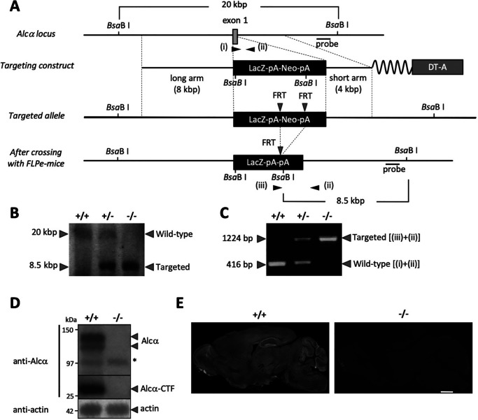
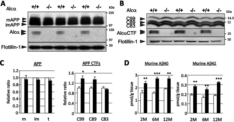
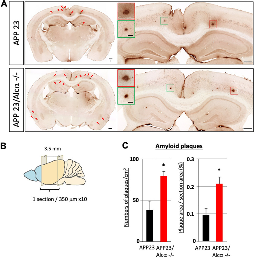
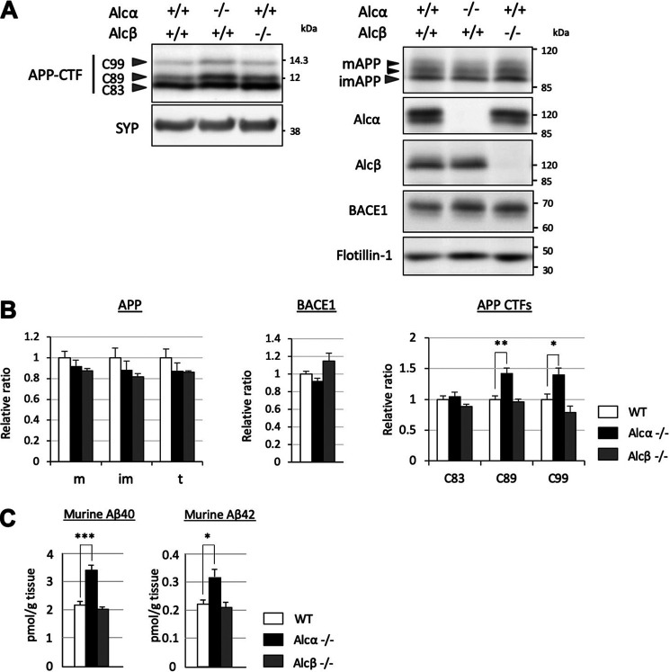
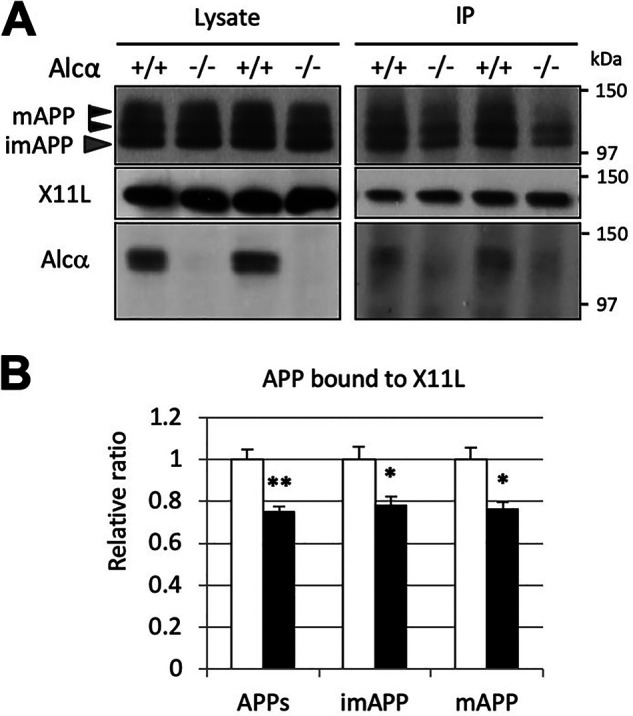
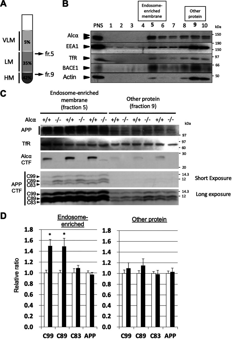
Alzheimer's disease (AD) is a very common neurodegenerative disorder, chiefly caused by increased production of neurotoxic β-amyloid (Aβ) peptide generated from proteolytic cleavage of β-amyloid protein precursor (APP). Except for familial AD arising from mutations in the APP and presenilin (PSEN) genes, the molecular mechanisms regulating the amyloidogenic processing of APP are largely unclear. Alcadein α/calsyntenin1 (ALCα/CLSTN1) is a neuronal type I transmembrane protein that forms a complex with APP, mediated by the neuronal adaptor protein X11-like (X11L or MINT2). Formation of the ALCα-X11L-APP tripartite complex suppresses Aβ generation in vitro, and X11L-deficient mice exhibit enhanced amyloidogenic processing of endogenous APP. However, the role of ALCα in APP metabolism in vivo remains unclear. Here, by generating ALCα-deficient mice and using immunohistochemistry, immunoblotting, and co-immunoprecipitation analyses, we verified the role of ALCα in the suppression of amyloidogenic processing of endogenous APP in vivo We observed that ALCα deficiency attenuates the association of X11L with APP, significantly enhances amyloidogenic β-site cleavage of APP, especially in endosomes, and increases the generation of endogenous Aβ in the brain. Furthermore, we noted amyloid plaque formation in the brains of human APP-transgenic mice in an ALCα-deficient background. These results unveil a potential role of ALCα in protecting cerebral neurons from Aβ-dependent pathogenicity in AD.

Keywords: Alzheimer's disease; Mint2; X11-like; alcadein; amyloid β (Aβ); amyloid β protein precursor (APP); calsyntenin; membrane protein; metabolism; neurodegeneration.


Conflict of interest statement

Conflict of interest—The authors declare that they have no conflicts of interest with the contents of this article.

Figures

Figure 1.
Figure 1.
Generation of Alcα-deficient mice. A, gene-targeting procedure. Shown is a schematic of the partial gene structure of the Alcα allele, including exon 1, the targeting construct, and targeted allele after crossing with FLPe-Tg mice. B, Southern blotting analysis. Probes indicated in A were used to detect WT (20 kbp) and the targeted (8.5 kbp) fragments. C, PCR products specific to the WT allele (+/+) generated with primers i plus ii (416 bp) and to the targeted allele (−/−) generated with primers ii plus iii (1,224 bp) were analyzed by agarose gel electrophoresis. D, immunoblot analysis of Alcα and Alcα CTF. Whole-brain lysates (20 μg of protein) of WT (+/+) and homozygous mutant (−/−) mice were analyzed in 8% resolving gel with an anti-Alcα antibody and anti-actin antibody. *, nonspecific product. E, immunostaining of sagittal sections of WT (+/+) and homozygous mutant (−/−) mouse (2–3 months old) brains with the anti-Alcα antibody. Scale bar, 1 mm.
Figure 2.
Figure 2.
Enhanced β-site cleavages of APP in the brains of Alcα-deficient mice. A and B, immunoblot analysis of APP (A) and APP CTFs (B). Membrane fraction (5 µg (A) or 15 µg (B) of protein) of the hippocampus and cerebral cortex of WT (+/+) and homozygous mutant (−/−) mice (3 months old) were analyzed in 8% (A) or 15% (B) resolving gel with anti-APP, anti-Alcα, and anti-flotillin-1 antibodies. APP, mature (N- and O-glycosylated APP695) and immature (N-glycosylated APP695) forms. Alcα CTF exhibits double bands under these electrophoresis conditions. C, band densities of APP and APP CTFs for WT (open columns) and Alcα-deficient (filled columns) mice were standardized to the density of flotillin-1, and the value of WT was assigned as a reference value of 1.0. m, mature APP (top two bands); im, immature APP (bottom band); t, total APP (mature plus immature APP); C99, CTFβ; C89, CTFβ′; C83, CTFα of APP CTFs (unpaired t test; *, p < 0.05; n = 3 mice/group). Error bars, S.E. D, endogenous mouse Aβ40 and Aβ42 in the hippocampus and cerebral cortex of WT (open columns) and Alcα-deficient (filled columns) mice at the indicated ages (2, 6, and 12 months old) were quantified using sandwich ELISA. The Aβ40 and Aβ42 concentrations were normalized to tissue weight (unpaired t test; **, p < 0.01; ***, p < 0.001; n = 5 mice/group). Error bars, S.E.
Figure 3.
Figure 3.
Quantification of amyloid plaques in APP23 mouse brain in the presence or absence of Alcα. A, immunostaining of coronal sections of brain regions including the cerebral cortex and hippocampus of APP23 (top) and APP23/Alcα-deficient mice (bottom) at 12 months of age. The brain sections were stained with anti-human Aβ antibody. Arrowheads, typical amyloid plaques. A magnified view of the area around the hippocampus and cortex is shown on the right with magnified images of plaques (squares in red and green). Scale bar, 300 μm (sections) or 50 μm (plaques). B and C, 10 35-µm-thick sections with 315-µm intervals were examined in one mouse. The total plaque numbers in 10 sections/mouse were counted and are indicated as the number (left) or area (right) of plaques per area (left) or section area (right). Error bars, S.E. (unpaired t test; *, p < 0.05; 4 mice for APP23, 3 mice for APP23/Alcα-deficient background).
Figure 4.
Figure 4.
Alcα-, but not Alcβ-deficient mice showed significant alterations in amyloidogenic processing of APP. A, immunoblot analysis of APP CTFs in Alcα- or Alcβ-deficient mice. A total of 15 µg of membrane fraction of the hippocampus and cerebral cortex of WT (+/+) and homozygous mutant (−/−) mice (6 months old) were analyzed in 15% resolving gel with anti-APP and anti-synaptophysin (SYP) antibodies. The same fractions were also analyzed in an 8% resolving gel with anti-Alcα, anti-Alcβ, anti-BACE1, and anti-flotillin-1 antibodies. B, band densities of APP CTFs for WT (black columns) and Alcα- or Alcβ-deficient (colored columns as indicated) were standardized to the density of synaptophysin, and the value of WT was assigned a reference value of 1.0. C99, CTFβ; C89, CTFβ′; C83, CTFα of APP CTFs (n = 4 mice/group; two-way ANOVA; Dunnett's post hoc test compared with WT; *, p < 0.05; **, p < 0.01). Band densities of APP (m, mature APP; im, immature APP; t, total APP) and BACE1 were also quantified and standardized to the density of flotillin-1, and the value of WT was assigned a reference value of 1.0. Error bars, S.E. C, endogenous mouse Aβ40 or Aβ42 in the hippocampus and cerebral cortex of WT (open column) and Alcα- or Alcβ-deficient (colored columns as indicated) mice were quantified using sandwich ELISA. The Aβ40 and Aβ42 concentrations were normalized to tissue weight (n = 4 mice/group; one-way ANOVA; Dunnett's post hoc test compared with WT; *, p < 0.05; ***, p < 0.001). Error bars, S.E.
Figure 5.
Figure 5.
Attenuated association of APP with X11L in the brains of Alcα-deficient mice. A, co-immunoprecipitation of APP with X11L in the presence or absence of Alcα. Crude membrane fractions (500 µg of protein) of the hippocampus and cerebral cortex of WT (+/+) and Alcα-deficient (−/−) mice were subject to immunoprecipitation with anti-X11L antibody, and the immunoprecipitates were analyzed by immunoblotting with anti-APP, anti-X11L, and anti-Alcα antibodies. B, the band densities of APP in A were quantified and standardized against X11L in the immunocomplex. APPs, mature APP plus immature APP; imAPP, immature APP; mAPP, mature APP. WT was assigned a reference value of 1.0. Statistical significance was analyzed using three independent experiments (n = 3 mice/group; unpaired t test; *, p < 0.05; **, p < 0.01). Data represent means ± S.E. (error bars).
Figure 6.
Figure 6.
Increased β-site cleavages of APP in the endosome-enriched fraction of Alcα-deficient mouse brains. A, preparation of endosome-enriched membrane fraction. Post-nuclear supernatant (PNS) prepared from brain homogenate was adjusted to 42.5% sucrose and set at the bottom. Buffer with 35% sucrose was overlaid, and the same buffer with 5% sucrose was subsequently applied as shown in the figure. Very light membrane (VLM) largely composed of late endosomes entered the 5% sucrose layer, and heavy membrane (HM)-containing plasma membrane and rough endoplasmic reticulum membrane with cytosol proteins resided in the 42.5% sucrose layer. Light membrane (LM)-containing early endosomes with Golgi and other membranes accumulated underneath the interface between 5 and 35% sucrose layers after ultracentrifugation. See Fig. S5 for briefly illustrated preparation scheme. B, typical isolation of endosome-enriched fraction from WT mouse hippocampus and cerebral cortex. The endosome-enriched fraction (fr. 5) and other protein fractions (fr. 9) are shown in boldface type. PNS and the respective fractions were analyzed by immunoblotting with antibodies to detect indicated proteins. Some EEA1 and BACE1 resided in the endosome-enriched fraction 5. ∼10% of the protein was separated in fraction 5, and ∼80% of protein resided in fractions 9 and 10 (Fig. S5). TfR, transferrin receptor. C, immunoblot analysis of the endosome-enriched (fr. 5) and other protein (fr. 9) fractions of WT and Alcα-deficient mouse brains. Samples of WT (+/+) and Alcα-deficient (−/−) mice were analyzed with antibodies to detect the indicated proteins. D, the band densities of APP and APP CTFs in C were quantified and standardized against transferrin receptor. The value of WT was assigned a reference value of 1.0. Statistical significance was analyzed using three independent experiments (n = 3 mice/group; unpaired t test; *, p < 0.05). Data represent means ± S.E. (error bars). APP, mature APP plus immature APP; C99, CTFβ; C89, CTFβ′; C83, CTFβ of APP CTFs.

Similar articles

Cited by

References

    1. Takami M., and Funamoto S. (2012) γ-Secretase-dependent proteolysis of transmembrane domain of amyloid precursor protein: successive tri- and tetrapeptide release in amyloid β-protein production. Int. J. Alzheimers Dis. 2012, 591392 10.1155/2012/591392 - DOI - PMC - PubMed
    1. Kakuda N., Shoji M., Arai H., Furukawa K., Ikeuchi T., Akazawa K., Takami M., Hatsuta H., Murayama S., Hashimoto Y., Miyajima M., Arai H., Nagashima Y., Yamaguchi H., Kuwano R., et al. (2012) Altered γ-secretase activity in mild cognitive impairment and Alzheimer's disease. EMBO Mol. Med. 4, 344–352 10.1002/emmm.201200214 - DOI - PMC - PubMed
    1. Forman M. S., Cook D. G., Leight S., Doms R. W., and Lee M.-Y. V. (1997) Differential effects of the Swedish mutant amyloid precursor protein on β-amyloid accumulation and secretion in neurons and nonneuronal cells. J. Biol. Chem. 272, 32247–32253 10.1074/jbc.272.51.32247 - DOI - PubMed
    1. Mullan M., Crawford F., Axelman K., Houlden H., Lilius L., Winblad B., and Lannfelt L. (1992) A pathogenic mutation for probable Alzheimer's disease in the APP gene at N-terminus of β-amyloid. Nat. Genet. 1, 345–347 10.1038/ng0892-345 - DOI - PubMed
    1. Goate A., Chartier-Harlin M.-C., Mullan M., Brown J., Crawford F., Fidani L., Giuffra L., Haynes A., Irving N., James L., Mant R., Newton P., Rooke K., Roques P., Talbot C., et al. (1991) Segregation of a missense mutation in the amyloid precursor protein gene with familial Alzheimer's disease. Nature 349, 704–706 10.1038/349704a0 - DOI - PubMed

Publication types

MeSH terms