. 2020 Sep;19(9):1468-1484.
doi: 10.1074/mcp.RA120.002078. Epub 2020 May 28.

Differential Complex Formation via Paralogs in the Human Sin3 Protein Interaction Network

Affiliations

Differential Complex Formation via Paralogs in the Human Sin3 Protein Interaction Network

Mark K Adams et al. Mol Cell Proteomics. 2020 Sep.

Abstract

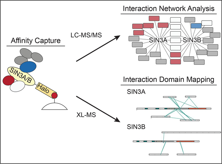
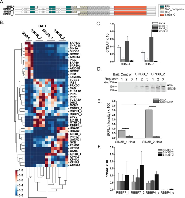
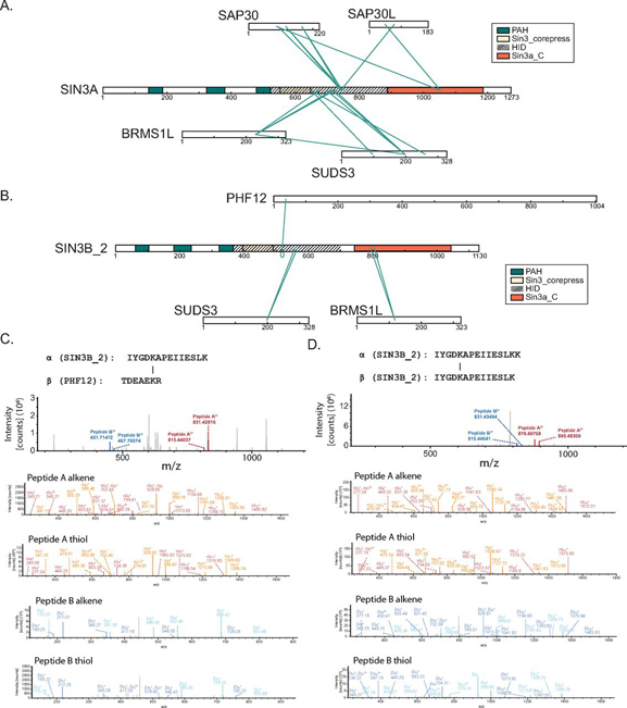
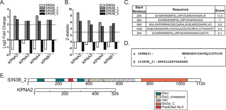
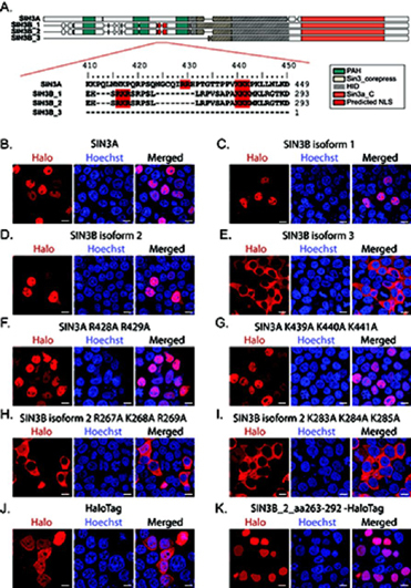
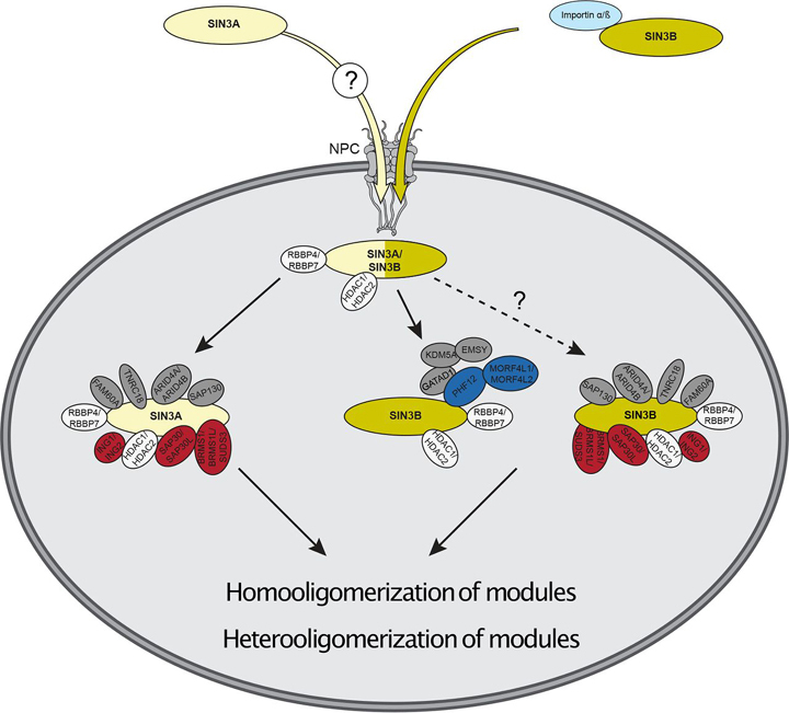
Despite the continued analysis of HDAC inhibitors in clinical trials, the heterogeneous nature of the protein complexes they target limits our understanding of the beneficial and off-target effects associated with their application. Among the many HDAC protein complexes found within the cell, Sin3 complexes are conserved from yeast to humans and likely play important roles as regulators of transcriptional activity. The presence of two Sin3 paralogs in humans, SIN3A and SIN3B, may result in a heterogeneous population of Sin3 complexes and contributes to our poor understanding of the functional attributes of these complexes. Here, we profile the interaction networks of SIN3A and SIN3B to gain insight into complex composition and organization. In accordance with existing data, we show that Sin3 paralog identity influences complex composition. Additionally, chemical cross-linking MS identifies domains that mediate interactions between Sin3 proteins and binding partners. The characterization of rare SIN3B proteoforms provides additional evidence for the existence of conserved and divergent elements within human Sin3 proteins. Together, these findings shed light on both the shared and divergent properties of human Sin3 proteins and highlight the heterogeneous nature of the complexes they organize.

Keywords: Chromatin function or biology; DSSO; SIN3; cross linking; epigenetics; histone deacetylase; nuclear translocation; pathway analysis; protein complex analysis; protein-protein interactions*; subcellular analysis; systems biology*.


Conflict of interest statement

Conflict of interest—Authors declare no competing interests.

Figures

None
Graphical abstract
Fig. 1
Fig. 1
Analysis of recombinant SIN3A and SIN3B_2 interaction networks.AB, Subcellular localization of stably expressed (A) SIN3A (NP_001138829.1) and (B) SIN3B_2 (NP_001284524.1) in Flp-In™-293 cells. HaloTag® TMRDirect™ Ligand and Hoechst 33258 solution were used to visualize recombinant protein localization (red) and nuclei (blue), respectively. White bars indicate 10 μm. CD, Plots of Z-statistic versus log2 fold change for the proteins detected in each APMS analysis of (C) SIN3A and (D) SIN3B_2 (supplemental Table S2D). Filter values used for enriched protein identification, Z-statistics ≥ 3 and log2 fold change values ≥ 2, are represented as dashed lines. E, Network of proteins with at least one isoform enriched by SIN3A (SIN3A-HaloTag) and/or SIN3B isoform 2 (SIN3B_2-HaloTag). Recombinant forms of SIN3A and SIN3B_2 are source nodes (yellow). Proteins with homology to Rpd3L-specific components (red), proteins with homology to Rpd3S-specific components (blue), and proteins with homology to proteins found within both Rpd3L and Rpd3S complexes (white) are displayed. Proteins with no clear homology to yeast Sin3 complex components (gray) are also shown. F, Spectrum matching a peptide specific to untagged SIN3A that was observed following SIN3A-HaloTag affinity purification.
Fig. 2
Fig. 2
Construction of the human Sin3 interaction network.A, Jaccard similarity indexes calculated using identities of enriched proteins within each bait protein purification were used as input for hierarchical clustering. Clustering was performed using the unweighted pair group method with arithmetic mean algorithm. Clustering of baits reveals 3 groups of proteins. B, Network of Halo-tagged bait proteins that enrich SIN3A and/or SIN3B. Halo-tagged bait proteins are target nodes and SIN3A/SIN3B are source nodes (yellow). Proteins with homology to Rpd3L-specific components (red), proteins with homology to Rpd3S-specific components (blue), and proteins with homology to proteins found within both Rpd3L and Rpd3S complexes (white) are displayed. Proteins with no clear homology to yeast Sin3 complex components (gray) are also shown. C, Bait normalized dNSAF (bdNSAF) of Rpd3S and Rpd3L component homologs with peptides observed following SIN3A-HaloTag and/or SIN3B_2-HaloTag affinity purification. D, bdNSAF of SIN3A and SIN3B following affinity purification of Rpd3S and Rpd3L component homologs expressed as fusions with HaloTag. CD, bdNSAF values were acquired by dividing prey protein dNSAF by bait protein dNSAF.
Fig. 3
Fig. 3
Chemical cross-linking MS analysis of SIN3A-HaloTag and SIN3B-HaloTag.AB, Connectivity maps showing cross-links involving proteins identified in our analysis as enriched by (A) SIN3A-HaloTag or (B) SIN3B_2-HaloTag (Fig. 1E, supplemental Tables S2E, S3). A–B, Proteins with homology to Rpd3L-specific components (red), proteins with homology to Rpd3S-specific components (blue), and proteins with homology to proteins found within both Rpd3L and Rpd3S complexes (white) are displayed. Proteins with no clear homology to yeast Sin3 complex components (gray) are also shown. CD, Maps of identified cross-links between (C) SIN3A-HaloTag or (D) SIN3B_2-HaloTag and the Sin3 complex components HDAC1/HDAC2 and RBBP4/RBBP7. PAH domains (green), Sin3_corepress domains (tan), and Sin3a_C domains (orange) are displayed. The experimentally defined SIN3A HID and a highly homologous region within SIN3B are designated by dashed lines.
Fig. 4
Fig. 4
Assessment of SIN3B domain organization with rare SIN3B isoforms.A, Visual alignment of human SIN3A (NP_001138829.1), SIN3B_1 (NP_056075.1), SIN3B_2 (NP_001284524.1), and SIN3B_3 (NP_001284526.1). PAH domains (green), Sin3_corepress domains (tan), and Sin3a_C domains (orange) are displayed. The experimentally defined SIN3A HID and a highly homologous region within SIN3B are designated by dashed lines. Detailed sequence alignments are provided in supplemental Fig. S2. B, Clustered heatmap of normalized dNSAF values for proteins within each bait purification replicate that were enriched by at least one Sin3 bait protein. Proteins were isolated and analyzed from three (SIN3A) or four (all SIN3B isoforms) replicates of cells stably expressing the recombinant Sin3 protein of interest (supplemental Table S4A–S4E). Values were standardized by subtracting the minimum value and dividing by the maximum values for each prey species. Clustering was performed using the unweighted pair group method with arithmetic mean algorithm. Proteins for which multiple isoforms are represented are denoted by isoform identifiers after the protein name. C, Average dNSAF values measured for HDAC1 and HDAC2 in SIN3A- (white), SIN3B_1 (light grey), SIN3B_2 (dark grey), and SIN3B_3 (black) affinity-purified samples. Mean ± S.D., n = 3 for SIN3A, and n = 4 for all SIN3B isoforms (supplemental Table S4E). DE, HaloTag (Control), SIN3B_1-HaloTag (SIN3B_1), SIN3B_2-HaloTag (SIN3B_2) were transiently expressed in 293T cells and purified using Magne® HaloTag® Beads. Purified protein was eluted in 100 µL of elution buffer. D, Ten µL of eluate from each transfection replicate was loaded onto 4–15% polyacrylamide gels and blots were probed with anti-SIN3B and goat-anti-mouse secondary antibody. E, HDAC activity assay of protein complexes purified using SIN3B_1 (SIN3B_1_Halo) and SIN3B_2 (SIN3B_2_Halo) transiently expressed within 293T cells as baits. Reactions were supplemented with DMSO (grey) or DMSO + SAHA (black). Relative fluorescence unit (RFU) values for all biological replicates were normalized to recombinant SIN3B protein abundance in purified samples as measured by Western blot (Fig. 3D, supplemental Fig. S7, supplemental Table S5). Mean ± S.D., n = 3. *: p ≤ 0.005, **: p ≤ 0.001. F, Average dNSAF values measured for RBBP7 and RBBP4 proteoforms in SIN3B_1 (light grey), SIN3B_2 (dark grey), and SIN3B_3 (black) affinity-purified samples. Mean ± S.D., n = 4 (supplemental Table S4E).
Fig. 5
Fig. 5
Identification of interfaces mediating interactions between Rpd3L/Rpd3S component homologs and SIN3A/SIN3B.AB, Map of identified cross-links between (A) SIN3A-HaloTag or (B) SIN3B-HaloTag and proteins with homology to Rpd3L- and Rpd3S-specific component homologs (supplemental Table S3). AB, PAH domains (green), Sin3_corepress domains (tan), and Sin3a_C domains (orange) are displayed. The experimentally defined SIN3A HID and a highly homologous region within SIN3B are designated by dashed lines. C, Peptides cross-linked to one another that map to SIN3B_2 and PHF12. MS2 spectrum (top spectrum) and MS3 spectra (bottom 4 spectra) are displayed. D, Peptides cross-linked to one another that represent a self-link between separate SIN3B molecules. MS2 spectrum (top spectrum) and MS3 spectra (bottom 4 spectra) are displayed.
Fig. 6
Fig. 6
Assessment of SIN3B interactions with karyophorin proteins.A, Log2 fold change and (B) Z-statistic values calculated by QSPEC v1.3.5 for KPNA2, KPNA3, KPNA4, and KPNB1 in SIN3A- (white), SIN3B_1- (light grey), SIN3B_2- (dark grey), and SIN3B_3- (black) purified samples (supplemental Table S4D). C, Potential SIN3B NLS signals identified by cNLS Mapper (41). D, Peptides cross-linked to one another that map to KPNA2 and SIN3B_2 (supplemental Table S3B). E, Map of identified cross-link between KPNA2 and SIN3B_2. SIN3B_2 PAH domains (green), the Sin3_corepress domain (tan), and the Sin3a_C domain (orange) are displayed. A region that aligns with the experimentally defined HID region within SIN3A is designated by dashed lines. A predicted NLS with a cNLS mapper score of 12.5 is displayed in red.
Fig. 7
Fig. 7
Characterization of the predicted SIN3B nuclear localization signal. (A, top panel) Alignment of the cNLS Mapper (41) predicted bipartite nuclear localization sequence in SIN3B (red). PAH domains (green), Sin3_corepress domains (tan), and Sin3a_C domains (orange) are displayed. The experimentally defined SIN3A HID and a highly homologous region within SIN3B are designated by dashed lines. (A, bottom panel) Residues mutated to test predicted NLS accuracy are highlighted in red. Alignment residue number (top) and sequence residue numbers (right) are displayed. BI, Subcellular localization of transiently expressed WT (BE) and mutant (FI) forms of Halo-tagged recombinant Sin3 proteoforms in 293T cells. FI, Protein names are appended with the identities of mutated residues. JK, Subcellular localization of transiently expressed HaloTag (J) and a sequence containing the predicted SIN3B NLS, spanning residues 263 to 292 of SIN3B_2, with a C-terminal HaloTag (K) in 293T cells. (BK) HaloTag® TMRDirect™ Ligand and Hoechst 33258 solution were used to visualize recombinant protein localization (red) and nuclei (blue), respectively. White bars indicate 10 μm.
Fig. 8
Fig. 8
Model of the human SIN3A and SIN3B interactions defined in this study. SIN3A and SIN3B have divergent nuclear localization signals that mediate their nuclear import. The SIN3B NLS serves as recognition signal for importin (light blue) and mediates nuclear import. PHF12 and known PHF12 interaction partners likely represent SIN3B-specific interaction partners. Homooligomerization and heterooligomerization of Sin3 modules may occur. NPC: nuclear pore complex. Rpd3L component homologs (red), Rpd3S component homologs (dark blue), Rdp3L/Rpd3S shared component homologs (white), and other known Sin3 interaction partners (gray) are displayed.

References

    1. Ibn-Salem J., Muro E.M., Andrade-Navarro M.A. Co-regulation of paralog genes in the three-dimensional chromatin architecture. Nucleic Acids Res. 2017;45:81–91. - PMC - PubMed
    1. Kondrashov F.A. Gene duplication as a mechanism of genomic adaptation to a changing environment. Proc. Biol. Sci. 2012;279:5048–5057. - PMC - PubMed
    1. Link S., Spitzer R.M.M., Sana M., Torrado M., Völker-Albert M.C., Keilhauer E.C., Burgold T., Pünzeler S., Low J.K.K., Lindström I., Nist A., Regnard C., Stiewe T., Hendrich B., Imhof A., Mann M., Mackay J.P., Bartkuhn M., Hake S.B. PWWP2A binds distinct chromatin moieties and interacts with an MTA1-specific core NuRD complex. Nat. Commun. 2018;9:4300. - PMC - PubMed
    1. Adams G.E., Chandru A., Cowley S.M. Co-repressor, co-activator and general transcription factor: the many faces of the Sin3 histone deacetylase (HDAC) complex. Biochem. J. 2018;475:3921–3932. - PMC - PubMed
    1. Carrozza M.J., Li B., Florens L., Suganuma T., Swanson S.K., Lee K.K., Shia W.J., Anderson S., Yates J., Washburn M.P., Workman J.L. Histone H3 methylation by Set2 directs deacetylation of coding regions by Rpd3S to suppress spurious intragenic transcription. Cell. 2005;123:581–592. - PubMed

Publication types

MeSH terms

LinkOut - more resources