Review
. 2020 May 28;147(10):dev175596.
doi: 10.1242/dev.175596.

The extracellular matrix in development

Affiliations
Review

The extracellular matrix in development

David A Cruz Walma et al. Development. .

Abstract

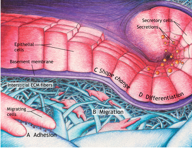
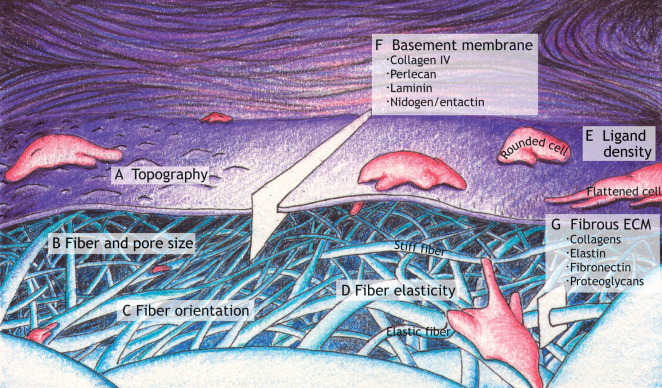
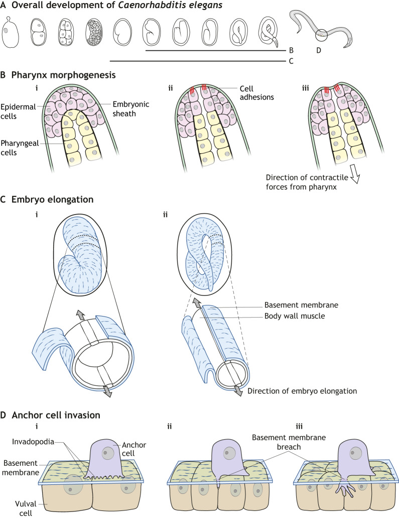
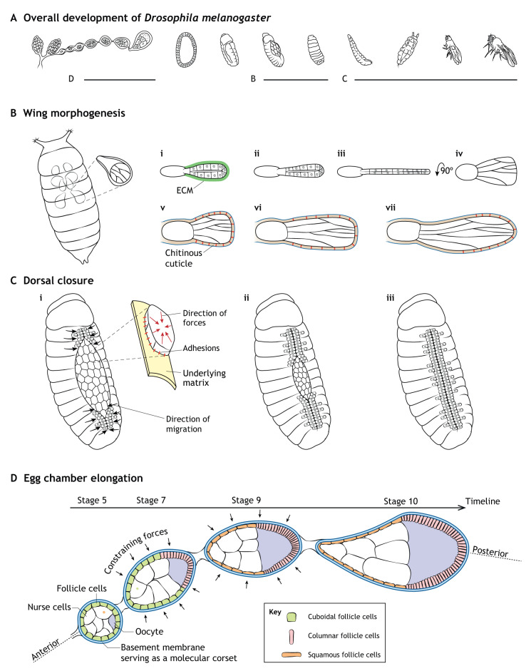
As the crucial non-cellular component of tissues, the extracellular matrix (ECM) provides both physical support and signaling regulation to cells. Some ECM molecules provide a fibrillar environment around cells, while others provide a sheet-like basement membrane scaffold beneath epithelial cells. In this Review, we focus on recent studies investigating the mechanical, biophysical and signaling cues provided to developing tissues by different types of ECM in a variety of developing organisms. In addition, we discuss how the ECM helps to regulate tissue morphology during embryonic development by governing key elements of cell shape, adhesion, migration and differentiation.

Keywords: Adhesion; Biophysical; Differentiation; Embryo; Extracellular matrix; Migration.


Conflict of interest statement

Competing interestsThe authors declare no competing or financial interests.

Figures

Fig. 1.
Fig. 1.
A summary of the regulation of developmental processes by the extracellular matrix. Two major forms of ECM are basement membranes and interstitial matrices. These types of ECM help to direct cell and tissue shape during morphogenesis in development by influencing cell adhesion (A), migration (B), morphology (C) and differentiation (D). (A) Cell-ECM adhesion, with cell adhesion complexes between the cell and a fibril substrate shown being mediated by integrins (heterodimeric receptors projecting downward from the closest migrating cell). (B) Migrating cells using oriented protrusions and cell-ECM adhesion complexes to move along interstitial ECM fibers. (C) Epithelial cells undergoing shape change (columnar-to-cuboidal transitions). (D) Epithelial cells differentiating into secretory cells.
Fig. 2.
Fig. 2.
Examples of physical properties of the extracellular matrix. (A) Topography encountered by a migrating cell. (B) Examples of varying fiber diameters and sizes of pores between ECM fibers. (C) Examples of fiber orientation: compare oriented fibers near ‘C’ with the other relatively non-oriented fibers. (D) Examples of varying fiber elasticity/stiffness represented as different degrees of fiber deformation as a cell pulls on two fibers using cell processes and cellular contractility. (E) Ligand density (shown as black bristles) affecting the extent of cell spreading. (F) Basement membrane composition: a slice of the basement membrane indicating key molecular components. (G) Fibrous ECM composition: a slice of fibrillar ECM listing several key components.
Fig. 3.
Fig. 3.
Schematic diagrams of C. elegans model systems discussed in this Review. (A) Overview of C. elegans development indicating stages involved in the following panels. (B) Pharynx morphogenesis. Epidermal cells adhering via cell adhesions to the surrounding embryonic sheath, which prevents deformation of the epidermis by pulling forces from the developing pharynx (pharyngeal cells in yellow). (C) Embryo elongation. The basement membrane serves as a ‘molecular corset’, acting in conjunction with muscle contractions to elongate the embryo. (D) Anchor cell invasion. Anchor cells use invadopodia to produce initial focal sites of basement membrane degradation (i). Upon breaching the basement membrane (ii), further invadopodia formation ceases, a large invasive protrusion forms and the anchor cell inserts itself between underlying vulval cells (iii).
Fig. 4.
Fig. 4.
Schematics of Drosophila model systems discussed in this Review. (A) Overview of Drosophila development indicating stages involved in the following panels. (B) Wing morphogenesis. (i-iv) Removal of the ECM initiates wing elongation secondary to cell columnar-to-cuboidal shape changes. (v-vii) Dynamic patterned attachment of pupal wing epithelial cells to the chitinous cuticle shapes the developing wing. (C) Early (i), middle (ii) and late (iii) dorsal closure. Contracting cells adhering to underlying matrix along with lateral epidermal cells migrating towards the dorsal midline as the amniosera contracts and ingresses. (D) Egg chamber elongation. The basement membrane promotes cuboidal (green)-to-squamous (orange) transitions of anterior follicle cells and cuboidal-to-columnar (pink) transitions of posterior follicle cells; the basement membrane provides constraining forces as a ‘molecular corset’ to elongate the egg chamber.

References

    1. Abagnale G., Steger M., Nguyen V. H., Hersch N., Sechi A., Joussen S., Denecke B., Merkel R., Hoffmann B., Dreser A. et al. (2015). Surface topography enhances differentiation of mesenchymal stem cells towards osteogenic and adipogenic lineages. Biomaterials 61, 316-326. 10.1016/j.biomaterials.2015.05.030 - DOI - PubMed
    1. Accogli A., Calabretta S., St-Onge J., Boudrahem-Addour N., Dionne-Laporte A., Joset P., Azzarello-Burri S., Rauch A., Krier J., Fieg E. et al. (2019). De Novo pathogenic variants in N-cadherin cause a syndromic neurodevelopmental disorder with corpus collosum, axon, cardiac, ocular, and genital defects. Am. J. Hum. Genet. 105, 854-868. 10.1016/j.ajhg.2019.09.005 - DOI - PMC - PubMed
    1. Afasizheva A., Devine A., Tillman H., Fung K. L., Vieira W. D., Blehm B. H., Kotobuki Y., Busby B., Chen E. I. and Tanner K. (2016). Mitogen-activated protein kinase signaling causes malignant melanoma cells to differentially alter extracellular matrix biosynthesis to promote cell survival. BMC Cancer 16, 186 10.1186/s12885-016-2211-7 - DOI - PMC - PubMed
    1. Alberts B., Johnson A., Lewis J., Morgan D., Raff M., Roberts K. and Walter P. (2014). Molecular Biology of the Cell. New York, NY: Garland Science.
    1. Alcaraz J., Otero J., Jorba I. and Navajas D. (2018). Bidirectional mechanobiology between cells and their local extracellular matrix probed by atomic force microscopy. Semin. Cell Dev. Biol. 73, 71-81. 10.1016/j.semcdb.2017.07.020 - DOI - PubMed

Publication types