

Maternal and fetal T cells in term pregnancy and preterm labor
- PMID: 32467619
- PMCID: PMC7331691
- DOI: 10.1038/s41423-020-0471-2
Maternal and fetal T cells in term pregnancy and preterm labor
Abstract
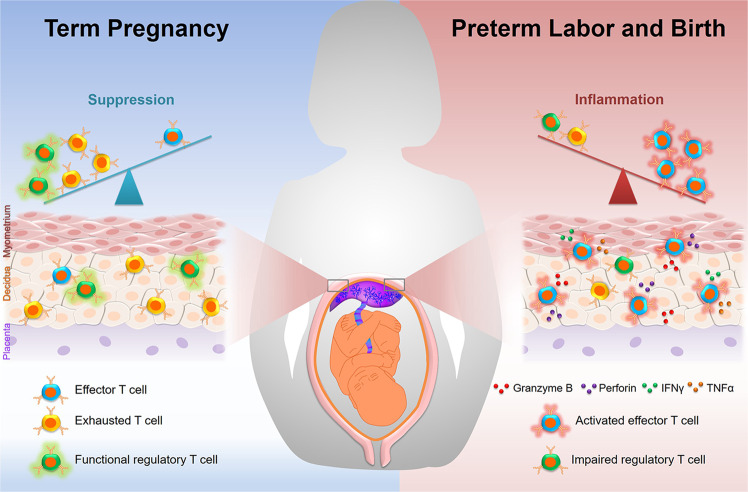
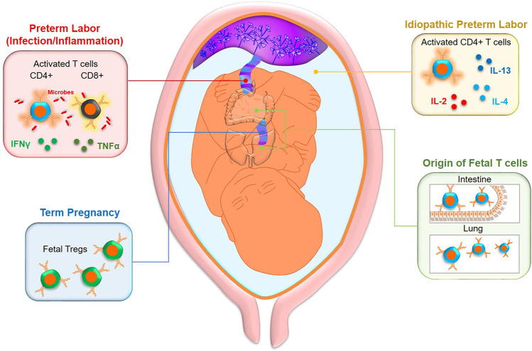
Pregnancy is a state of immunological balance during which the mother and the developing fetus must tolerate each other while maintaining sufficient immunocompetence to ward off potential threats. The site of closest contact between the mother and fetus is the decidua, which represents the maternal-fetal interface. Many of the immune cell subsets present at the maternal-fetal interface have been well described; however, the importance of the maternal T cells in this compartment during late gestation and its complications, such as preterm labor and birth, has only recently been established. Moreover, pioneer and recent studies have indicated that fetal T cells are activated in different subsets of preterm labor and may elicit distinct inflammatory responses in the amniotic cavity, leading to preterm birth. In this review, we describe the established and proposed roles for maternal T cells at the maternal-fetal interface in normal term parturition, as well as the demonstrated contributions of such cells to the pathological process of preterm labor and birth. We also summarize the current knowledge of and proposed roles for fetal T cells in the pathophysiology of the preterm labor syndrome. It is our hope that this review provides a solid conceptual framework highlighting the importance of maternal and fetal T cells in late gestation and catalyzes new research questions that can further scientific understanding of these cells and their role in preterm labor and birth, the leading cause of neonatal mortality and morbidity worldwide.
Keywords: adaptive immunity; amniotic fluid; decidua; maternal-fetal interface; parturition.
Conflict of interest statement
The authors declare no competing interests.
Figures
References
-
- Bonney EA, Onyekwuluje J. The H-Y response in mid-gestation and long after delivery in mice primed before pregnancy. Immunol. Invest. 2003;32:71–81. - PubMed
-
- Aluvihare VR, Kallikourdis M, Betz AG. Regulatory T cells mediate maternal tolerance to the fetus. Nat. Immunol. 2004;5:266–271. - PubMed
-
- Robertson SA, Guerin LR, Moldenhauer LM, Hayball JD. Activating T regulatory cells for tolerance in early pregnancy—the contribution of seminal fluid. J. Reprod. Immunol. 2009;83:109–116. - PubMed
Publication types
MeSH terms
Grants and funding
LinkOut - more resources
Full Text Sources
