. 2020 Aug;44(2):637-649.
doi: 10.3892/or.2020.7615. Epub 2020 May 19.

Girdin interaction with vimentin induces EMT and promotes the growth and metastasis of pancreatic ductal adenocarcinoma

Affiliations

Girdin interaction with vimentin induces EMT and promotes the growth and metastasis of pancreatic ductal adenocarcinoma

Wulin Wang et al. Oncol Rep. 2020 Aug.

Abstract

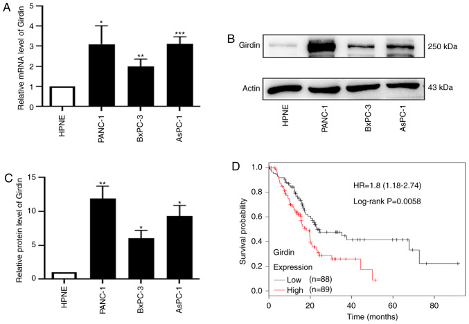
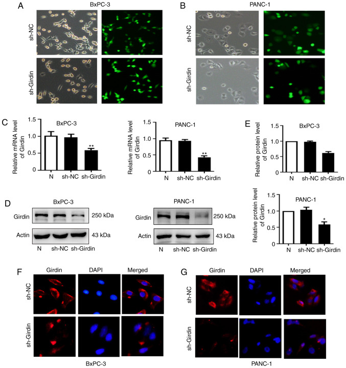
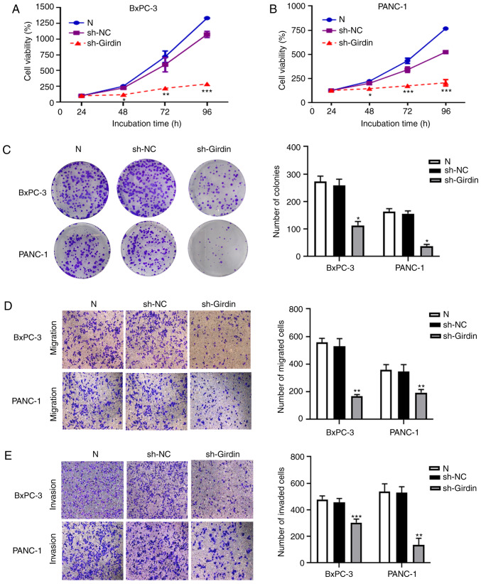
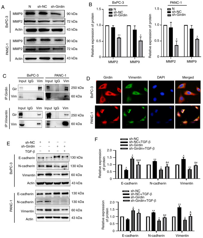
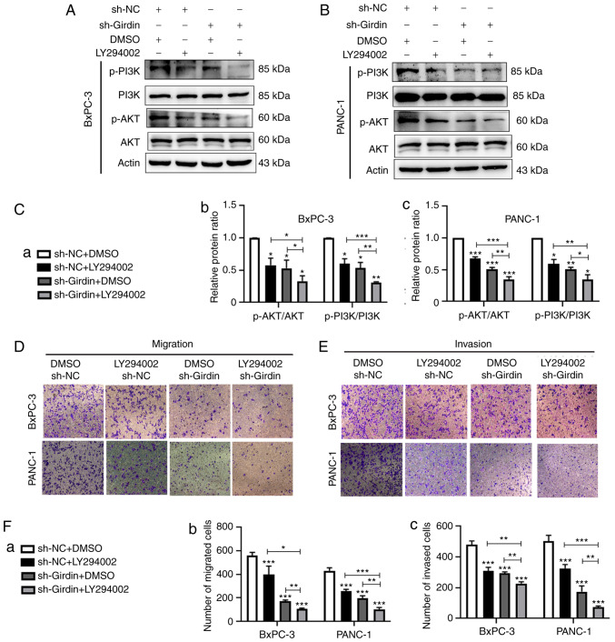
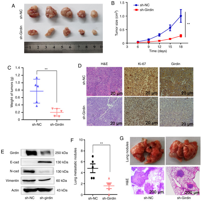
Pancreatic ductal adenocarcinoma (PDAC) is a highly malignant cancer of the digestive tract that has a high potential for metastasis and a poor prognosis. Girdin was first reported in 2005 as an actin‑binding protein and was designated as Akt‑phosphorylation enhancer (APE); thus, Girdin has been revealed to have an important role in regulating cancer development. There is additional evidence indicating that Girdin is associated with cell proliferation, migration, invasion and survival in certain cancers. However, the potential mechanisms involving Girdin and mobility in pancreatic cancer have not been elucidated. In the present study, it was revealed that Girdin was highly expressed in pancreatic cancer tissue and was associated with tumor grade. The present study, to the best of our knowledge, is the first aimed at investigating the unknown role of Girdin in PDAC metastasis. A short hairpin RNA for Girdin (sh‑Girdin) was successfully constructed with recombinant adenoviral vectors to suppress the expression of Girdin in pancreatic cancer cell lines (PANC‑1 and BXPC‑3). The silencing efficiency of the Girdin shRNA was determined by RT‑qPCR and western blot analysis, and decreased Girdin expression in the cytoplasm was revealed by immunofluorescence detection. Then, sulforhodamine B (SRB) and colony formation assays were used to confirm that the knockdown of Girdin inhibited proliferation in vitro, and Transwell assays were used to examine the influence of Girdin knockdown on cellular mobility. Animal experiments also confirmed that silencing the expression of Girdin in pancreatic cancer cells inhibited the growth and metastasis of pancreatic cancer in vivo. Transforming growth factor‑β (TGF‑β) is a common inducer of epithelial‑mesenchymal transition (EMT) and can effectively induce EMT in PDAC. Notably, TGF‑β‑treated cells exhibited changes in the classic biological markers of EMT. The expression of E‑cadherin, a marker of the epithelial phenotype, increased, and the expression of N‑cadherin and vimentin, markers of the interstitial phenotype, decreased in response to sh‑Girdin. According to these experiments, Girdin may affect pancreatic cancer progression and development by interacting with vimentin. Therefore, there is evidence indicating that Girdin could be designated as a prognostic biological indicator and a candidate therapeutic target for pancreatic cancer.

Keywords: Girdin; growth; metastasis; pancreatic ductal adenocarcinoma; EMT.


Figures

Figure 1.
Figure 1.
Girdin is upregulated in PDAC, and high expression of Girdin is associated with poor survival. (A) Relative mRNA expression of Girdin in three PDAC cell lines, PANC-1, BxPC-3 and AsPC-1, compared with the normal human pancreatic ductal cell line hTERT-HPNE was assessed by RT-qPCR. (B) Protein levels of Girdin in the PDAC cell lines PANC-1, BxPC-3 and AsPC-1 and the normal human pancreatic ductal cell line hTERT-HPNE were confirmed by western blotting. (C) Histogram of the protein expression of Girdin in the PDAC cell lines PANC-1, BxPC-3 and AsPC-1 and the normal human pancreatic ductal cell line hTERT-HPNE. (D) Kaplan-Meier plotter analysis of GEPIA(http://gepia.cancer -pku.cn/) data indicated that a high level of Girdin was associated with poor survival; high (n=88) and low (n=89). Error bars were expressed as the mean ± SD, and the experiment was performed at least in triplicate. *P<0.05, **P<0.01, and ***P<0.001 compared to HPNE. PDAC, pancreatic ductal adenocarcinoma; RT-qPCR, real-time quantitative polymerase chain reaction; Actin, internal reference; GEPIA, Gene Expression Profiling Interactive Analysis.
Figure 2.
Figure 2.
Girdin gene expression is decreased by a specific sh-RNA (sh-Girdin). (A and B) Fluorescence microscopy image with a scale bar of 100 µm to confirm the transfection efficiency, indicating successful infection by sh-NC and sh-Girdin in the PANC-1 and BxPC-3 cells. (C) Real-time quantitative PCR was used to evaluate the mRNA level of Girdin in PDAC cell lines transfected with recombinant adenovirus. Actin was used as an internal control. (D and E) Girdin expression in BxPC-3 and PANC-1 cells was assessed by western blotting, and the protein level of Girdin was significantly decreased in the sh-Girdin groups. (F and G) PANC-1 and BxPC-3 cells were transfected with sh-NC or sh-Girdin and analyzed for protein expression and cellular distribution by immunofluorescence staining with an antibody against Girdin (red), showing that the cytoplasmic staining of Girdin was suppressed by sh-Girdin. Nuclei were stained and labeled with DAPI (blue). Scale bars, 100 µm. Significant results were obtained from at least three independent experiments; the data are presented as the mean ± SD. *P<0.05, and **P<0.01 compared with sh-NC. PDAC, pancreatic ductal adenocarcinoma; sh-NC, non-targeting control short hairpin RNA; sh-Girdin, a specific short hairpin RNA of Girdin.
Figure 3.
Figure 3.
Effects of sh-Girdin on the proliferation, migration and invasion of pancreatic cancer cells. (A) The absorbance value of control cells and BxPC-3 cells transfected with sh-Girdin incubated for various time periods (24, 48,72 and 96 h) was measured by SRB assay. Knockdown of Girdin significantly reduced proliferation after incubation for 48 h. (B) The cell viability of BxPC-3 cells was measured by SRB assay at an absorbance of 490 nm. The proliferation of PANC-1 cells was inhibited by sh-Girdin. (C) The number of colonies was used to investigate proliferation after the cells were treated for 14 days, and knockdown of Girdin suppressed the formation of colonies in PDAC. (D and E) Transwell assays were used to confirm the metastasis of PDAC cells in vitro. Treatment with sh-Girdin reduced the number of migrating and invading cells in the lower chambers, scale bar, 100 µm. Three experiments were independently performed. The data are presented as the mean ± SD. *P<0.05, **P<0.01, and ***P<0.001 compared with sh-NC. PDAC, pancreatic ductal adenocarcinoma; SRB, sulforhodamine B; sh-NC, non-targeting control short hairpin RNA; sh-Girdin, a specific short hairpin RNA of Girdin.
Figure 4.
Figure 4.
Girdin interacts with vimentin to reverse TGF-β-induced EMT. (A and B) Girdin was knocked down by sh-Girdin, and the cells were treated for 48 h; western blotting was used to analyze the protein levels of MMP2 and MMP9 in the PDAC cell lines. (C) Girdin-interacting proteins were assessed in PANC-1 and BxPC-3 cells by co-IP, which revealed the endogenous Girdin-vimentin interaction. The lysates were analyzed by western blotting. (D) Endogenous Girdin and vimentin in PANC-1 and BxPC-3 cells was detected by immunofluorescence staining. The cells were not treated before fixation with 4% paraformaldehyde, and the cellular location of Girdin (red) and vimentin (green) was detected by immunofluorescence staining using anti-Girdin and anti-vimentin antibodies. The nuclei were stained with DAPI (blue) and visualized by fluorescence microscopy at a scale bar of 100 µm. (E and F) The EMT markers E-cadherin, N-cadherin and vimentin were evaluated by western blotting. As revealed, sh-Girdin reduced the protein levels of N-cadherin and vimentin compared with those in the sh-NC group, and the TGF-β-induced change in the expression of these markers was reversed by sh-Girdin. Three experiments were independently performed. The data are presented as the mean ± SD. *P<0.05, and **P<0.01 compared with sh-NC; &P<0.05, compared with sh-NC co-incubated with TGF-β; #P<0.05, and ##P<0.01 compared with sh-Girdin. PDAC, pancreatic ductal adenocarcinoma; Gir, Girdin; Vim, Vimentin; TGF-β, transforming growth factor-β; EMT, epithelial-mesenchymal transition; sh-NC, non-targeting control short hairpin RNA; sh-Girdin, a specific short hairpin RNA of Girdin.
Figure 5.
Figure 5.
Effects of Girdin knockdown on the inhibition of metastasis in PDAC are enhanced by LY294002. (A and B) Western blot analysis of the level of phosphorylation of PI3K and Akt expression in PANC-1 and BxPC-3 cells. (C) Statistical analysis of the ratio of phosphorylated protein to total protein. Knockdown of Girdin inactivated phosphorylation of the PI3K/AKT pathway. LY294002 enhanced the inhibitory effect of Girdin knockdown on PDAC. (D and E) The migration and invasion abilities of PDAC cells were evaluated by Transwell assay. (F) Statistical analysis of PDAC cell migration and invasion; scale bar, 100 µm. Three experiments were independently performed. The data are presented as the mean ± SD. *P<0.05, **P<0.01, and ***P<0.001. PDAC, pancreatic ductal adenocarcinoma; DMSO, dimethyl sulfoxide; sh-NC, non-targeting control short hairpin RNA; sh-Girdin, a specific short hairpin RNA of the Girdin.
Figure 6.
Figure 6.
Girdin regulates PDAC cell tumor growth and metastasis in vivo. (A) Stable Girdin-knockdown BxPC-3 cells were injected under the skin of nude mice. (B) Tumor volumes were first calculated after injection for 6 days and measured every three days after. (C) The tumors were dissected and measured, and the tumor weights in the sh-Girdin group were reduced compared to those in the sh-NC group. (D) Immunostaining revealed H&E staining and staining using antibodies against Ki-67 and Girdin in xenograft tumors. The levels of Ki-67 and Girdin in the samples from the sh-Girdin group were lower than those in the control group. Scale bar, 20 µm. (E) The protein level of Girdin was also detected by western blotting, and the changes in EMT markers were similar to those observed in vitro. (F) Stable Girdin-knockdown BxPC-3 cells were injected into the mice through the tail vein, and six weeks later, the lung metastasis in the sh-Girdin group was significantly decreased compared with that in the sh-NC group based on the number of nodules and the area of H&E staining. Scale bar, 200 µm. The tumor weight and the lung metastatic nodules are presented as the mean ± SD. **P<0.01 compared with sh-NC group. PDAC, pancreatic ductal adenocarcinoma; H&E, hematoxylin and eosin; EMT, epithelial-mesenchymal transition; sh-NC, non-targeting control short hairpin RNA; sh-Girdin, a specific short hairpin RNA of Girdin.

Similar articles

Cited by

References

    1. Owens DK, Davidson KW, Krist AH, Barry MJ, Cabana M, Caughey AB, Curry SJ, Doubeni CA, Epling JW, Kubik M, et al. Screening for pancreatic cancer. JAMA. 2019;322:438. doi: 10.1001/jama.2019.10232. - DOI - PubMed
    1. Bray F, Ferlay J, Soerjomataram I, Siegel RL, Torre LA, Jemal A. Global cancer statistics 2018: GLOBOCAN estimates of incidence and mortality worldwide for 36 cancers in 185 countries. CA Cancer J Clin. 2018;68:394–424. doi: 10.3322/caac.21492. - DOI - PubMed
    1. Moffat GT, Epstein AS, O'Reilly EM. Pancreatic cancer-A disease in need: Optimizing and integrating supportive care. Cancer. 2019;125:3927–3935. doi: 10.1002/cncr.32423. - DOI - PMC - PubMed
    1. Rahib L, Fleshman JM, Matrisian LM, Berlin JD. Evaluation of pancreatic cancer clinical trials and benchmarks for clinically meaningful future trials: A systematic review. JAMA Oncol. 2016;2:1209–1216. doi: 10.1001/jamaoncol.2016.0585. - DOI - PubMed
    1. Rahib L, Smith BD, Aizenberg R, Rosenzweig AB, Fleshman JM, Matrisian LM. Projecting cancer incidence and deaths to 2030: The unexpected burden of thyroid, liver, and pancreas cancers in the United States. Cancer Res. 2014;74:2913–2921. doi: 10.1158/0008-5472.CAN-14-0155. - DOI - PubMed

MeSH terms