. 2020 Aug;46(2):773-781.
doi: 10.3892/ijmm.2020.4617. Epub 2020 May 27.

Hyperoxia reduces STX17 expression and inhibits the autophagic flux in alveolar type II epithelial cells in newborn rats

Affiliations

Hyperoxia reduces STX17 expression and inhibits the autophagic flux in alveolar type II epithelial cells in newborn rats

Dan Zhang et al. Int J Mol Med. 2020 Aug.

Abstract

Supplemental oxygen therapy can be life‑saving for premature infants. Our previous study revealed a defect in the autophagic flux in the lung tissues of neonatal rats with hyperoxia‑induced bronchopulmonary dysplasia (BPD), but the underlying mechanism remains unknown. Moreover, there are few innovative treatments that can completely alter the course of BPD. The present study examined the expression of Syntaxin 17 (STX17), a protein necessary for autophagosome‑lysosome binding, in alveolar type II (AT‑II) epithelial cells of neonatal rats with BPD. Neonatal Sprague‑Dawley rats were randomly exposed to elevated O2 levels [fraction of inspired oxygen (FiO2), 0.8; model group] or normal room air (FiO2, 0.21; control group), and the expression levels of STX17, autophagy‑related [Microtubule‑associated protein 1A/1B‑light chain 3B (LC3B)‑II, p62, lysosomal‑associated membrane protein 1)] and apoptosis‑related (cleaved caspase3) mRNA and proteins were examined in lung tissues. Moreover, the expression levels of the aforementioned proteins were measured in isolated primary AT‑II cells cultured in vitro under hyperoxic conditions in the presence or absence of pharmacological modulators of autophagy. Transmission electron microscopy identified that AT‑II cell apoptosis and autophagosome aggregation were elevated in the lungs of BPD rats compared with control rats on postnatal day 7. STX17 mRNA and protein expression levels were decreased in lung tissue and isolated AT‑II cells as early as postnatal day 3 in BPD rats, while the expression levels of LC3B‑II, p62 and cleaved caspase3 were increased, reaching a peak on postnatal day 7. This early reduction in STX17 expression, followed by increased expression in autophagy‑ and apoptosis‑related proteins, was also observed in isolated AT‑II cells exposed to hyperoxia in vitro. However, treatment with the autophagy inducers rapamycin or LiCl eliminated the hyperoxia‑induced reduction in STX17, partially restored the autophagy flux and increased the survival of AT‑II cells exposed to hyperoxia. Collectively, these results indicated that STX17 expression in AT‑II cells was reduced in the early stages of BPD in neonatal rats and may be related to the subsequent hyperoxia‑induced block in autophagic flux.


Figures

Figure 1
Figure 1
Structure of lung tissues from rats with hyperoxia-induced BPD. (A) Hematoxylin and eosin-stained lung sections from rats in the control and model groups on postnatal days 1, 3, 7 and 14. Magnification, ×200. (B) Transmission electron microscopy of alveolar type II cells from rats in the control and model groups on postnatal day 7. White arrow indicates autophagosome aggregation. Magnification, ×20,000 (local, x2).
Figure 2
Figure 2
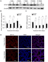
STX17 protein and mRNA expression levels in the lungs of BPD rats. (A) Western blot analysis of STX17 expression in the lung tissues of rats in the control group and BPD model group on the indicated days after birth and (B) semi-quantification of the results. (C) Reverse transcription-quantitative PCR analysis of STX17 mRNA expression in the lung tissues of rats in control and model group. (D) Immunofluorescence staining of STX17 protein in the lung tissues of rats in the control and model group (local, x2). Scale bar, 50 µm. *P<0.05, **P<0.01. C, control; M, model; BPD, bronchopulmonary dysplasia; STX17, syntaxin 17.
Figure 3
Figure 3
Characterization of primary AT-II cells. (A) Immunofluorescence staining of pro-SPC, an AT-II cell-specific marker, indicating that ≥90% of the cells were positive (red; magnification, ×200). (B) Light microscopy of AT-II cells showing paving stone-like changes (magnification, ×200). (C) Transmission electron microscopy images showing lamellar bodies in the cells (white arrow). Pro-SPC, surfactant; AT-II, alveolar type II (magnification, ×20,000).
Figure 4
Figure 4
Expression of STX17 and autophagy- and apoptosis-related proteins in hyperoxic AT-II cells. (A) Western blot analysis of the (B) autophagy-related proteins LC3B-II, (C) p62 and (D) Lamp1, the apoptosis-related protein (E) cleaved caspase3 and (F) STX17 in primary AT-II cells. *P<0.05, **P<0.01. AT-II, alveolar type II; STX17, syntaxin 17; LC3B, Microtubule-associated protein 1A/1B-light chain 3B; Lamp1, Lysosomal-associated membrane protein 1; C, control; M, model.
Figure 5
Figure 5
Expression of STX17 and autophagy- and apoptosis-related mRNAs in hyperoxia AT-II cells. RT-qPCR analysis of (A) LC3B, (B) p62, (C) Lamp1 and (D) STX17 mRNA expression levels in primary AT-II cells. **P<0.01. AT-II, alveolar type II; STX17, syntaxin 17; LC3B, Microtubule-associated protein 1A/1B-light chain 3B; Lamp1, Lysosomal-associated membrane protein 1.
Figure 6
Figure 6
Expression of STX17 and autophagy- and apoptosis-related proteins in primary AT-II cells exposed to hyperoxia. (A) Western blot analysis of LC3B-II, p62 and STX17 in AT-II cells exposed to hyperoxia for the indicated times. (B) MTT proliferation assay of primary AT-II cells incubated with RAPA, LiCl, 3-MA and/or CQ. (C) Western blot analysis of LC3B-II and cleaved caspase3 in AT-II cells. (D) Western blot analysis of STX17 expression in AT-II cells incubated in the presence or absence of RAPA, LiCl or CQ. RAPA, rapamycin; 3-MA, 3-methyladenine; CQ, chloroquine; M, model; AT-II, alveolar type II; STX17, syntaxin 17; LC3B, Microtubule-associated protein 1A/1B-light chain 3B; Lamp1, Lysosomal-associated membrane protein 1; OD, optical density.

Similar articles

Cited by

References

    1. Thébaud B, Goss KN, Laughon M, Whitsett JA, Abman SH, Steinhorn RH, Aschner JL, Davis PG, McGrath-Morrow SA, Soll RF, Jobe AH. Bronchopulmonary dysplasia. Nat Rev Dis Primers. 2019;5:78. doi: 10.1038/s41572-019-0127-7. - DOI - PMC - PubMed
    1. Stoll BJ, Hansen NI, Bell EF, Shankaran S, Laptook AR, Walsh MC, Hale EC, Newman NS, Schibler K, Carlo WA, et al. Neonatal outcomes of extremely preterm infants from the NICHD Neonatal research network. Pediatrics. 2010;126:443–456. doi: 10.1542/peds.2009-2959. - DOI - PMC - PubMed
    1. Jobe AH. The new bronchopulmonary dysplasia. Curr Opin Pediatr. 2011;23:167–172. doi: 10.1097/MOP.0b013e3283423e6b. - DOI - PMC - PubMed
    1. Kinsella JP, Greenough A, Abman SH. Bronchopulmonary dysplasia. Lancet. 2006;367:1421–1431. doi: 10.1016/S0140-6736(06)68615-7. - DOI - PubMed
    1. Rock JR, Hogan BL. Epithelial progenitor cells in lung development, maintenance, repair, and disease. Annu Rev Cell Dev Biol. 2011;27:493–512. doi: 10.1146/annurev-cellbio-100109-104040. - DOI - PubMed

MeSH terms

Substances