. 2020 Aug;46(2):561-570.
doi: 10.3892/ijmm.2020.4610. Epub 2020 May 19.

MicroRNA‑93 contributes to the suppression of lung inflammatory responses in LPS‑induced acute lung injury in mice via the TLR4/MyD88/NF‑κB signaling pathway

Affiliations

MicroRNA‑93 contributes to the suppression of lung inflammatory responses in LPS‑induced acute lung injury in mice via the TLR4/MyD88/NF‑κB signaling pathway

Hu Gao et al. Int J Mol Med. 2020 Aug.

Abstract

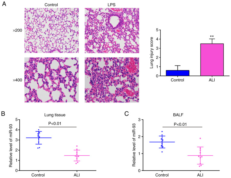
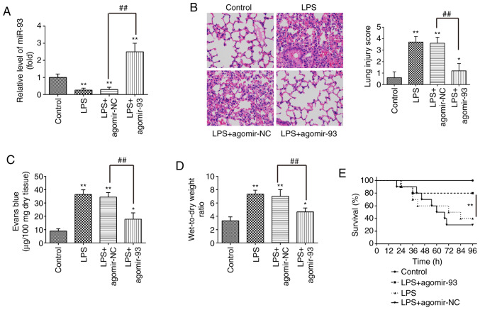
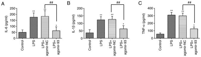
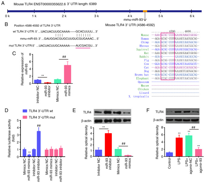
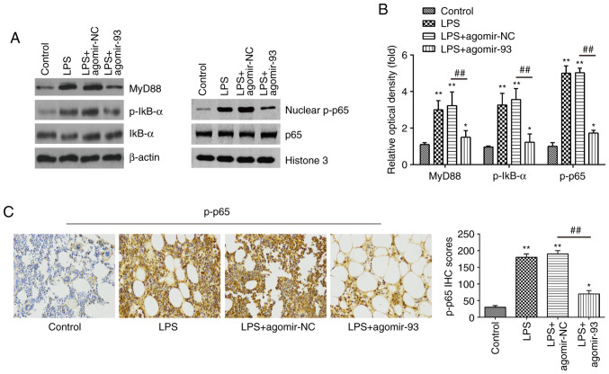
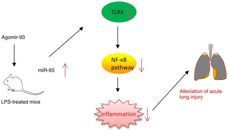
Acute lung injury (ALI) is a severe inflammatory lung disease with a rapid onset. The anti‑inflammatory functions of microRNA‑93 (miRNA/miR‑93) have been described in various types of tissue injury and disease. However, the biological role of miR‑93 and its molecular mechanisms underlying the initiation and progression of ALI have not yet been reported, at least to the best of our knowledge. The present study aimed to investigate the regulatory effects exerted by miR‑93 in ALI. Using an in vivo murine model of ALI induced by lipopolysaccharide (LPS), miR‑93 expression was found to be downregulated in the lung tissues and bronchoalveolar lavage fluid (BALF) compared with the control group. Following agomiR‑93 injection, it was observed that agomiR‑93 attenuated lung injury, as evidenced by decreased lung permeability, a reduced lung wet/dry weight ratio and an increased survival rate of the mice. Concomitantly, agomiR‑93 significantly reduced LPS‑induced the interleukin (IL)‑6, IL‑1β, and tumor necrosis factor (TNF)‑α levels in BALF. Of note, Toll‑like receptor 4 (TLR4), an upstream regulator of the nuclear factor (NF)‑κB signaling pathway, was directly suppressed by miR‑93 in RAW 264.7 cells. Importantly, agomiR‑93 induced a significant suppression of the TLR4/myeloid differentiation primary response 88 (MyD88)/NF‑κB signaling pathway, as demonstrated by the downregulation of MyD88, and the phosphorylation of IκB‑α and p65 in the lung tissues of mice with ALI. Taken together, the findings of the present study indicate that miR‑93 attenutes LPS‑induced lung injury by regulating the TLR4/MyD88/NF‑κB signaling pathway, suggesting that miR‑93 may prove to be a potential therapeutic target for ALI.


Figures

Figure 1
Figure 1
miR-93 is downregulated in mice with LPS-induced ALI. Mice (n=6 in each group) were treated with a single dose of LPS (2 mg/kg) or normal saline and total RNA was isolated from their lungs 24 h following treatment. (A) Staining images of lung tissues following hematoxylin and eosin staining and the pathological changes in lung tissues were evaluated semi-quantitatively based on a histological examination. Original magnification, ×200 and ×400. Data are the means ± SD (n=3) of one representative experiment, **P<0.01 vs. the control group. (B and C) miR-93 expression was determined by RT-qPCR in lung tissues and BALF of mice challenged with LPS (n=10). P<0.01 vs. the control group. LPS, lipopolysaccharide; ALI, acute lung injury.
Figure 2
Figure 2
miR-93 attenuates lung injury in mice with LPS-induced ALI. Mice were injected intravenously with agomir-93 and control agomir (8 mg/kg) 24 h prior to the LPS (2 mg/kg) challenge. Following the LPS administration for 24 h, the mice were sacrificed and the lung tissues were collected for analysis. (A) miR-93 expression was assessed in lung tissues in the model of ALI induced by LPS (n=6 in each group). (B) Staining images of lung tissues following hematoxylin and eosin staining and the pathological changes in lung tissues were evaluated semi-quantitatively based on a histological examination (n=6 in each group). Original magnification, ×100. (C) Lung permeability was assessed using the Evans blue dye extravasation method (n=6 in each group). (D) The lung W/D ratio was determined to evaluate pulmonary edema at 24 h after the LPS challenge (n=6 in each group). Data are the means ± SD (n=3) of one representative experiment, *P<0.05, **P<0.01 vs. the control group. ##P<0.01 vs. the LPS + agomir-NC group. (E) Survival rates were determined using the Kaplan-Meier method. **P<0.01 vs. the LPS alone group. LPS, lipopolysaccharide; ALI, acute lung injury; W/D, wet/dry ratio.
Figure 3
Figure 3
miR-93 suppresses the inflammatory response in mice with LPS-induced ALI. Mice were injected intravenously with agomir-93 and control agomir (8 mg/kg) 24 h prior to the LPS (2 mg/kg) challenge. After the LPS administration for 24 h, the mice were sacrificed and BALF was collected for analysis. (A) IL-6, (B) IL-1β, and (C) TNF-α levels in BALF were measured using commercial ELISA kits. Data are the means ± SD (n=3) of one representative experiment, *P<0.05, **P<0.01 vs. the control group. ##P<0.01 vs. the LPS + agomir-NC group. LPS alone group. LPS, lipopolysaccharide; ALI, acute lung injury; BALF, bronchoalveolar lavage fluid.
Figure 4
Figure 4
TLR4 is a direct target of miR-93. (A and B) The putative binding site of miR-93 and TLR4 is shown. (C) The expression levels of miR-93 were measured by RT-qPCR following transfection with miR-93 mimics or miR-93 inhibitor in RAW264.7 cells. (D) Luciferase activity of RAW264.7 cells was detected by a dual luciferase assay. Data are the means ± SD (n=3) of one representative experiment, **P<0.01 vs. mimics NC, ##P<0.01 vs. inhibitor NC. (E) Protein expression levels of TLR4 following transfection with miR-93 mimics or miR-93 inhibitor measured by western blot analysis. Data are the means ± SD (n=3) of one representative experiment, **P<0.01 vs mimics NC, ##P<0.01 vs inhibitor NC. (F) Mice were injected intravenously with agomir-93, and agomir NC (8 mg/kg) 24 h prior to the LPS (2 mg/kg) challenge. After the LPS administration for 24 h, the mice were sacrificed and lung tissues were then collected for the detection of the protein expression levels of TLR4 by western blot analysis. Data are the means ± SD (n=3) of one representative experiment, **P<0.01 vs. control group. ##P<0.01 vs. the LPS + agomiR-NC group. LPS, lipopolysaccharide; ALI, acute lung injury; TLR4, Toll-like receptor 4.
Figure 5
Figure 5
miR-93 inhibits the LPS-induced inflammatory response through the TLR4/MyD88/NF-κB pathway. Mice were injected intravenously with agomir-93, and agomir NC (8 mg/kg) 24 h prior to the LPS (2 mg/kg) challenge. After the LPS administration for 24 h, the mice were sacrificed and lung tissues were then collected for analysis. (A) The levels of MyD88, IκB-α, p-IκB-α, p65 and nuclear p-p65 were measured by western blot analysis. (B) The bands were semi-quantitatively analyzed using Image J software, and normalized to β-actin density. (C) The expression of nuclear p-p65 was determined by IHC (×400 magnification) and IHC scores are calculated. Data are the means ± SD (n=3) of one representative experiment. *P<0.05, **P<0.01 vs. control group. ##P<0.01 vs. LPS + agomiR-NC group. LPS, lipopolysaccharide; ALI, acute lung injury; TLR4, Toll-like receptor 4; MyD88, myeloid differentiation primary response 88.
Figure 6
Figure 6
Schematic diagram of the signaling pathway in which miR-93 attenuates LPS-induced acute lung injury. AgomiR-93 injection attenuated LPS-induced inflammation in a model of ALI by blocking the activation of the TLR4/MyD88/NF-κB signaling pathway. LPS, lipopolysaccharide; ALI, acute lung injury; TLR4, Toll-like receptor 4; MyD88, myeloid differentiation primary response 88.

Similar articles

Cited by

References

    1. Herridge MS, Tansey CM, Matte A, Tomlinson G, Diaz-Granados N, Cooper A, Guest CB, Mazer CD, Mehta S, Stewart TE, et al. Functional disability 5 years after acute respi-ratory distress syndrome. N Engl J Med. 2011;364:1293–1304. doi: 10.1056/NEJMoa1011802. - DOI - PubMed
    1. Mendez JL, Hubmayr RD. New insights into the pathology of acute respiratory failure. Curr Opin Crit Care. 2005;11:29–36. doi: 10.1097/00075198-200502000-00005. - DOI - PubMed
    1. Zhang J, Cao J, Feng J, Wu Q, Chen BY. A study of noninvasive positive-pressure mechanical ventilation in the treatment of acute lung injury with a complex critical care ventilator. J Int Med Res. 2014;42:788–798. doi: 10.1177/0300060514522205. - DOI - PubMed
    1. Tsushima K, King LS, Aggarwal NR, De Gorordo A, D'Alessio FR, Kubo K. Acute lung injury review. Intern Med. 2009;48:621–630. doi: 10.2169/internalmedicine.48.1741. - DOI - PubMed
    1. Deng J, Wang DX, Liang AL, Tang J, Xiang DK. Effects of baicalin on alveolar fluid clearance and α-ENaC expression in rats with LPS-induced acute lung injury. Can J Physiol Pharmacol. 2017;95:122–128. doi: 10.1139/cjpp-2016-0212. - DOI - PubMed

MeSH terms