

Early detection of ovarian cancer
- PMID: 32468105
- PMCID: PMC7476911
- DOI: 10.1007/s00330-020-06937-z
Early detection of ovarian cancer
Abstract
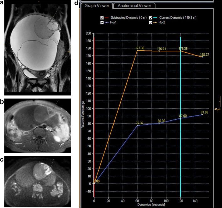
Early detection is the only way to achieve a high cure rate in women with ovarian cancer. Unfortunately, to date, there is no effective strategy for early detection, despite rapidly emerging biomarkers. The low prevalence of ovarian cancer, low specificity and high rates of false positives have been limitations of screening programs. In the hands of experts, transvaginal sonography and MRI are effective tools to characterise ovarian masses. Currently, ongoing efforts in standardization of technique and analysis are likely to improve diagnostic capabilities in clinical routine, as well as the introduction of predictive risk models of malignancy. Radiomics and radiogenomics potentially offer a broad spectrum of complementary information in ovarian cancer diagnosis and treatment. KEY POINTS: • Transvaginal sonography and MRI are effective tools to characterise ovarian masses. • Standardisation of imaging technique and implementation of predictive models of risk of malignancy contribute to early detection of ovarian cancer.
Keywords: Epithelial ovarian cancer; Magnetic resonance imaging; Ovarian cancer; Radiomics; Screening.
Conflict of interest statement
The author of this manuscript declares no relationships with any companies, whose products or services may be related to the subject matter of the article.
Figures
References
-
- Nougaret S, Tardieu M, Vargas HA et al (2019) Ovarian cancer: an update in the area of radiomics. Diagn Interv Imaging 100:647–655 - PubMed
-
- Carlson KJ (2020) Screening for ovarian cancer www.uptodate.com
-
- Thomassin-Naggara I, Poncelet E, Jalaguier-Coudray A, et al. Ovarian-adnexal reporting data system magnetic resonance imaging (O-RADS MRI) score for risk stratification of sonographically indeterminate adnexal masses. JAMA Netw Open. 2020;3:e1919896. doi: 10.1001/jamanetworkopen.2019.19896. - DOI - PMC - PubMed
Publication types
MeSH terms
LinkOut - more resources
Full Text Sources
Medical
