Spectrum of Cardiac Manifestations in COVID-19: A Systematic Echocardiographic Study
- PMID: 32469253
- PMCID: PMC7382541
- DOI: 10.1161/CIRCULATIONAHA.120.047971
Spectrum of Cardiac Manifestations in COVID-19: A Systematic Echocardiographic Study
Abstract
Background: Information on the cardiac manifestations of coronavirus disease 2019 (COVID-19) is scarce. We performed a systematic and comprehensive echocardiographic evaluation of consecutive patients hospitalized with COVID-19 infection.
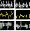
Methods: One hundred consecutive patients diagnosed with COVID-19 infection underwent complete echocardiographic evaluation within 24 hours of admission and were compared with reference values. Echocardiographic studies included left ventricular (LV) systolic and diastolic function and valve hemodynamics and right ventricular (RV) assessment, as well as lung ultrasound. A second examination was performed in case of clinical deterioration.
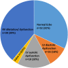
Results: Thirty-two patients (32%) had a normal echocardiogram at baseline. The most common cardiac pathology was RV dilatation and dysfunction (observed in 39% of patients), followed by LV diastolic dysfunction (16%) and LV systolic dysfunction (10%). Patients with elevated troponin (20%) or worse clinical condition did not demonstrate any significant difference in LV systolic function compared with patients with normal troponin or better clinical condition, but they had worse RV function. Clinical deterioration occurred in 20% of patients. In these patients, the most common echocardiographic abnormality at follow-up was RV function deterioration (12 patients), followed by LV systolic and diastolic deterioration (in 5 patients). Femoral deep vein thrombosis was diagnosed in 5 of 12 patients with RV failure.
Conclusions: In COVID-19 infection, LV systolic function is preserved in the majority of patients, but LV diastolic function and RV function are impaired. Elevated troponin and poorer clinical grade are associated with worse RV function. In patients presenting with clinical deterioration at follow-up, acute RV dysfunction, with or without deep vein thrombosis, is more common, but acute LV systolic dysfunction was noted in ≈20%.
Keywords: COVID-19; echocardiography; heart ventricles; thromboembolism.
Figures


Comment in
-
Letter by Vazgiourakis et al Regarding Article, "Spectrum of Cardiac Manifestations in COVID-19: A Systematic Echocardiographic Study".Circulation. 2021 Mar 2;143(9):e749-e750. doi: 10.1161/CIRCULATIONAHA.120.049322. Epub 2021 Mar 1. Circulation. 2021. PMID: 33646831 Free PMC article. No abstract available.
-
Letter by Carrizales-Sepúlveda et al Regarding Article, "Spectrum of Cardiac Manifestations in COVID-19: A Systematic Echocardiographic Study".Circulation. 2021 Mar 2;143(9):e751-e752. doi: 10.1161/CIRCULATIONAHA.120.049435. Epub 2021 Mar 1. Circulation. 2021. PMID: 33646833 Free PMC article. No abstract available.
References
-
- Lang RM, Badano LP, Mor-Avi V, Afilalo J, Armstrong A, Ernande L, Flachskampf FA, Foster E, Goldstein SA, Kuznetsova T, et al. Recommendations for cardiac chamber quantification by echocardiography in adults: an update from the American Society of Echocardiography and the European Association of Cardiovascular Imaging. J Am Soc Echocardiogr 2015281–39.e14doi: 10.1016/j.echo.2014.10.003 - PubMed
-
- Yancy CW, Jessup M, Bozkurt B, Butler J, Casey DE, Jr, Drazner MH, Fonarow GC, Geraci SA, Horwich T, Januzzi JL, et al. 2013 ACCF/AHA guideline for the management of heart failure: executive summary: a report of the American College of Cardiology Foundation/American Heart Association Task Force on Practice Guidelines. Circulation 20131281810–1852doi: 10.1161/CIR.0b013e31829e8807 - PubMed
MeSH terms
Substances
LinkOut - more resources
Full Text Sources
Other Literature Sources
Medical

