

Chest X-ray features of SARS-CoV-2 in the emergency department: a multicenter experience from northern Italian hospitals
- PMID: 32469732
- PMCID: PMC7243792
- DOI: 10.1016/j.rmed.2020.106036
Chest X-ray features of SARS-CoV-2 in the emergency department: a multicenter experience from northern Italian hospitals
Abstract
Objectives: To evaluate the imaging features of routine admission chest X-ray in patients referred for novel Coronavirus 2019 infection.
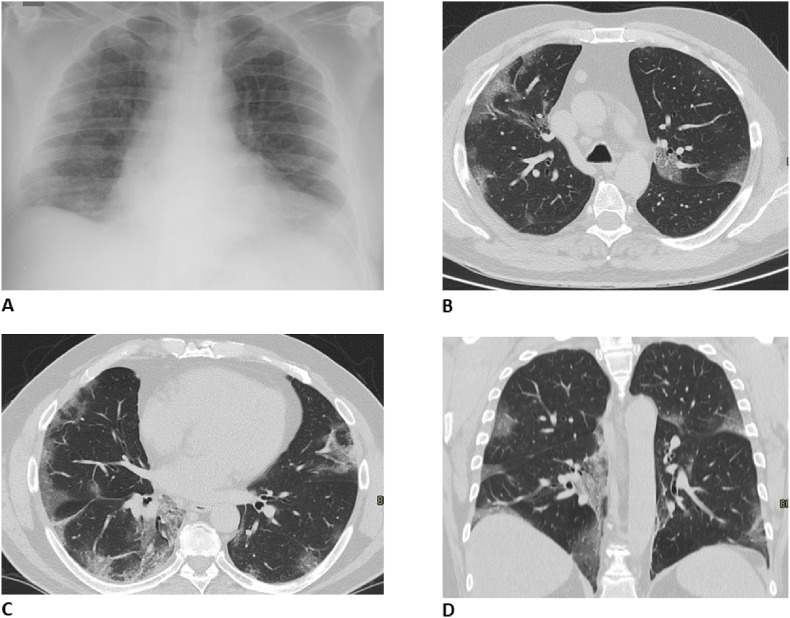
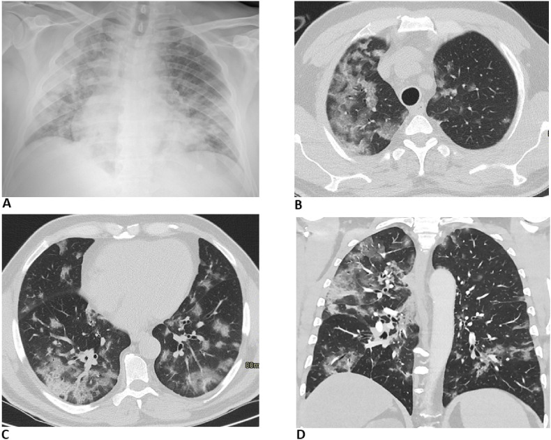
Methods: All patients referred to the emergency departments, RT-PCR positive for SARS-CoV-2 infection were evaluated. Demographic and clinical data were recorded. Two radiologists (8 and 15 years of experience) reviewed all the X-ray images and evaluated the following findings: interstitial opacities, alveolar opacities (AO), AO associated with consolidation, consolidation and/or pleural effusion. We stratified patients in groups according to the time interval between symptoms onset (cut-off 5 days) and X-ray imaging and according to age (cut-off 60 years old). Computed tomography was performed in case of a discrepancy between clinical symptoms, laboratory and X-ray findings, and/or suspicion of complications.
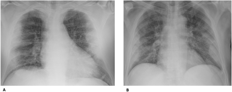
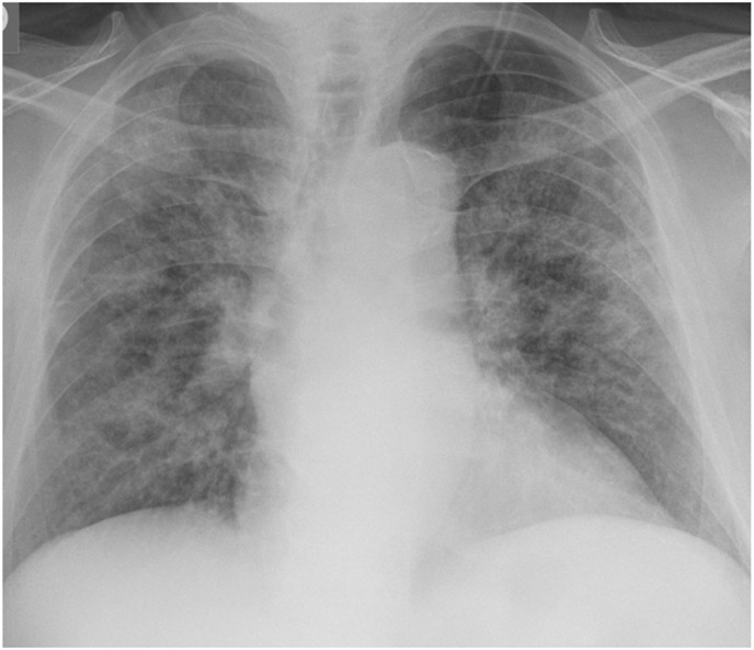
Results: A total of 468 patients were tested positive for SARS-CoV-2. Lung lesions primarily manifested as interstitial opacities (71.7%) and AO opacities (60.5%), more frequently bilateral (64.5%) and with a peripheral predominance (62.5%). Patients admitted to the emergency radiology department after 5 days from symptoms onset, more frequently had interstitial and AO opacities, in comparison to those admitted within 5 days, and lung lesions were more frequently bilateral and peripheral. Older patients more frequently presented interstitial and AO opacities in comparison to younger ones. Sixty-eight patients underwent CT that principally showed the presence of ground-glass opacities and consolidations.
Conclusions: The most common X-ray pattern is multifocal and peripheral, associated with interstitial and alveolar opacities. Chest X-ray, compared to CT, can be considered a reliable diagnostic tool, especially in the Emergency setting.
Keywords: Coronavirus; Infections; Radiography; Tomography; X-ray computed.
Copyright © 2020 Elsevier Ltd. All rights reserved.
Conflict of interest statement
The authors declare that they have no known competing financial interests or personal relationships that could have appeared to influence the work reported in this paper.
Figures

References
-
- Hui J.Y.-H., Hon T.Y.-W., Yang M.K.-W., et al. High-resolution computed tomography is useful for early diagnosis of severe acute respiratory syndrome–associated coronavirus pneumonia in patients with normal chest radiographs. J. Comput. Assist. Tomogr. 2004;28:1–9. doi: 10.1097/00004728-200401000-00001. - DOI - PubMed
Publication types
MeSH terms
LinkOut - more resources
Full Text Sources
Medical
Miscellaneous
