. 2020 May 29;15(5):e0233689.
doi: 10.1371/journal.pone.0233689. eCollection 2020.

The crystal structures of Thermus thermophilus CMP kinase complexed with a phosphoryl group acceptor and donor

Affiliations

The crystal structures of Thermus thermophilus CMP kinase complexed with a phosphoryl group acceptor and donor

Ryosuke Mega et al. PLoS One. .

Abstract

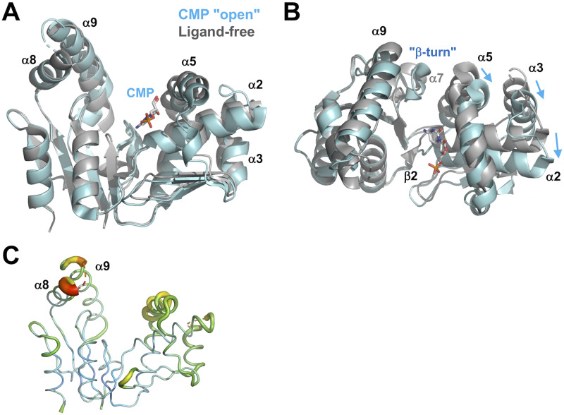
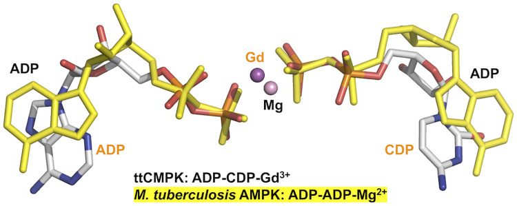
Nucleoside monophosphate kinases play crucial roles in biosynthesis and regeneration of nucleotides. These are bi-substrate enzymes that catalyze reversible transfers of a phosphoryl group between ATP and nucleoside monophosphate. These enzymes are comprised of the CORE domain, the NMP-binding domain, and the LID domain. Large conformational rearrangement of the three domains occurs during the catalytic cycle. Although many structures of CMP kinase have been determined, only limited structural information has been available on the conformational changes along the reaction pathway. We determined five crystal structures of CMP kinase of Thermus thermophilus HB8 in ligand-free form and the CMP "open", CMP "closed", ADP-CDP-Gd3+-, and CDP-bound forms at resolutions of 1.7, 2.2, 1.5, 1.6, and 1.7 Å, respectively. The ligand-free form was in an open conformation, whereas the structures of the CMP "closed", ADP-CDP-Gd3+-, and CDP-bound forms were in a closed conformation, in which the shift of the NMP-binding domain and LID domain caused closure of the substrate-binding cleft. Interestingly, the CMP "open" form was in an open conformation even with CMP bound, implying intrinsic conformational fluctuation. The structure of the ADP-CDP complex is the first structure of CMP kinase with a phosphoryl group donor and an acceptor. Upon simultaneous binding of ADP and CDP, the side chains of several residues in the LID domain moved toward the nucleotides without global open-closed conformational changes compared to those in the CMP "closed" and CDP complexes. These global and local conformational changes may be crucial for the substrate recognition and catalysis. The terminal phosphate groups of ADP and CDP had similar geometry to those of two ADP in AMP kinase, suggesting common catalytic mechanisms to other nucleoside monophosphate kinases. Our findings are expected to contribute to detailed understanding of the reaction mechanism of CMP kinase.


Conflict of interest statement

The authors have declared that no competing interests exist.

Figures

Fig 1
Fig 1. Crystal structure of ttCMPK.
(A) Overall structure of ttCMPK in complex with ADP, CDP, and Gd3+. The structure is shown in a schematic representation, colored in a spectrum from the N-terminus (blue) to the C-terminus (red). The bound nucleotides are shown in sticks. (B) Superimposition of the overall structure of ligand-free form (gray; PDB code 3W90), CMP "open" complex (cyan; 3W8N), CMP "closed" complex (green; 3AKE), ADP-CDP-Gd3+ complex (orange; 3AKC), and CDP complex (olive; 3AKD).
Fig 2
Fig 2. A schematic representation of the reaction cycle of ttCMPK.
The series of the conformational changes which were determined in this study (gray) or hypothesized (white) to occur upon substrate binding are shown. “A”, “C”, and “P” represent adenosine, cytidine, and phosphate moieties, respectively. The symbol ‡ indicates the transition state of phosphoryl transfer.
Fig 3
Fig 3. Superimposition of the structures of the ligand-free form (gray) and the CMP "open" complex (cyan).
(A) The two structures are superimposed on the basis of Cα positions in the CORE domain. CMP is shown as sticks colored according to atom type. Disordered regions are shown as dotted lines. (B) Compared to (A), this figure is rotated by 90° around the horizontal axis. (C) B-factor diagram of the ligand-free form represented by the B-factor putty program in PyMOL. The B-factor values are illustrated by color, ranging from low (blue) to high (red). The average B factor is 21.5 Å2.
Fig 4
Fig 4. Structural comparison of the CMP "closed" complex (green) with the ligand-free form (gray).
(A) The structures are superimposed on the basis of Cα positions in the CORE domain. CMP is shown as sticks colored according to atom type. Disordered regions are shown as dotted lines. (B) Compared to (A), this figure is rotated by 125° around the horizontal axis.
Fig 5
Fig 5. Structural comparison of the CMP "closed" complex (green) with the CMP "open" complex (cyan).
(A) The structures are superimposed on the basis of Cα positions in the CORE domain. CMP is shown as sticks colored according to atom type. Disordered regions are shown as dotted lines. (B) The structures are superimposed on the basis of bound CMP. (C) Interaction between the NMP-binding and LID domains in the CMP "closed" complex. Compared to panel B, this figure is rotated by 80° around the horizontal axis. The side chains are shown as sticks. Numbers are the distances shown in Å.
Fig 6
Fig 6. Interaction with CMP in the CMP "open" complex (A) and the CMP "closed" complex (B).
CMP is shown as sticks colored according to atom type. The side chains are shown as sticks. Numbers are the distances shown in Å.
Fig 7
Fig 7. Structural comparison of the ADP-CDP-Gd3+ (orange) complex with the CMP "closed" complex (green).
(A) The structures are superimposed on the basis of Cα positions in the whole proteins. ADP and CDP are shown as sticks colored according to atom type. The Gd3+ ion is shown as magenta sphere. Disordered regions are shown as dotted lines. (B) Compared to (A), this figure is rotated by 125° around the horizontal axis. (C) Recognition of the adenine ring of ADP.
Fig 8
Fig 8. Active site in the ADP-CDP-Gd3+ complex.
(A) Binding site of ADP and CDP. Possible interactions are shown as dashed lines. Numbers are the distances shown in Å. The Gd3+ ion is shown as magenta sphere. (B) Superimposition of the active site of the ADP-CDP-Gd3+ complex (light orange) with that of the CMP "closed" complex (light green). Note that the side chain of Leu195 is omitted from panel B because they occupy the position of the adenine ring of ADP (see Fig 7C). Superscripts L and N denote the residues from the LID and NMP-binding domains. Asterisks represent the residues whose main-chain amide groups are involved in ligand recognition. Arrows represent shifts of the side chains.
Fig 9
Fig 9. Coordination of the Gd3+ ion in the ADP-CDP-Gd3+ complex.
Oxygen atoms of water molecules and Gd3+ ion are shown as red and magenta spheres, respectively. Possible interactions are shown as dashed lines. Numbers are the distances shown in Å.
Fig 10
Fig 10. Structure of the CDP complex.
(A) The structures of the CDP complex (olive) and ADP-CDP-Gd3+ complex (orange) are superimposed on the basis of Cα positions in the whole proteins. ADP and CDP are shown as sticks. The side chains of Leu195 are shown as sticks. (B) Recognition of CDP. CDP and selected residues are colored as follows: olive, CDP complex; light orange, ADP-CDP-Gd3+ complex; and light green, CMP "closed" complex. The CDP and residues from the CDP complex are shown as thick sticks. Numbers are the distances shown in Å.
Fig 11
Fig 11. Comparison of CMPK structures.
(A) Ligand-free forms of CMPK from T. thermophilus (gray; PDB code 3W90), E. coli (magenta; 1CKE), and S. pneumoniae (brown; 1Q3T). (B) CMP-bound forms of CMPK from T. thermophilus (green; 3AKE), E. coli (teal; 1KDO), and S. aureus (light purple; 2H92). (C) CDP-bound forms of CMPK from T. thermophilus (olive; 3AKD) and E. coli (light brown; 2CMK). (D) CMP-bound forms of CMPK from T. thermophilus (green; 3AKE) and M. abscessus (light green; 4DIE), and ligand-free form of CMPK from M. abscessus (dark gray; 3R8C). These structures were structurally aligned based on superimposing the backbone structure of their CORE domains. For ttCMPK, the CMP "closed" complex was employed as CMP-bound form.
Fig 12
Fig 12. Comparison of CMP-binding sites between ttCMPK and E. coli CMPK.
The side chains are colored as follows: the CMP complex of E. coli CMPK (in dark green), the CMP "closed" complex of ttCMPK (in green), and the ADP-CDP-Gd3+ complex of ttCMPK (in orange). The names of residues in the ADP-CDP-Gd3+ complex are represented in parentheses. Arrows represent shifts of the side chains in the CMP "closed" complex to the ADP-CDP-Gd3+ complex of ttCMPK.
Fig 13
Fig 13. Bound nucleotides in the ternary complexes of ttCMPK (white) and Mycobacterium tuberculosis AMPK (yellow).
The nucleotides bound to ttCMPK are shown in the CPK model. Gd3+ and Mg2+ ions are in ttCMPK and Mycobacterium AMPK, respectively. The PDB codes for these structures are 3AKC (this study) and 2CDN [43].

Similar articles

Cited by

References

    1. Yan H, Tsai M. Nucleoside monophosphate kinases: structure, mechanism, and substrate specificity. Adv Enzymol Relat Areas Mol Biol. 1999; 73: 103–134. 10.1002/9780470123195.ch4 . - DOI - PubMed
    1. Vonrhein C, Schlauderer GJ, Schulz GE. Movie of the structural changes during a catalytic cycle of nucleoside monophosphate kinases. Structure. 1995; 3: 483–490. 10.1016/S0969-2126(01)00181-2 . - DOI - PubMed
    1. Müller C, Schulz G. Structure of the complex between adenylate kinase from Escherichia coli and the inhibitor Ap5A refined at 1.9 Å resolution. A model for a catalytic transition state. J Mol Biol. 1992; 224: 159–177. 10.1016/0022-2836(92)90582-5 . - DOI - PubMed
    1. Müller C, Schlauderer G, Reinstein J, Schulz G. Adenylate kinase motions during catalysis: an energetic counterweight balancing substrate binding. Structure. 1996; 4: 147–156. 10.1016/S0969-2126(96)00018-4 . - DOI - PubMed
    1. Henzler-Wildman K, Thai V, Lei M, Ott M, Wolf-Watz M, Fenn T, et al. Intrinsic motions along an enzymatic reaction trajectory. Nature. 2007; 450: 838–844. 10.1038/nature06410 . - DOI - PubMed

Publication types

MeSH terms