Review
. 2020 May 29;477(10):1983-2006.
doi: 10.1042/BCJ20200194.

The thick waxy coat of mycobacteria, a protective layer against antibiotics and the host's immune system

Affiliations
Review

The thick waxy coat of mycobacteria, a protective layer against antibiotics and the host's immune system

Sarah M Batt et al. Biochem J. .

Abstract

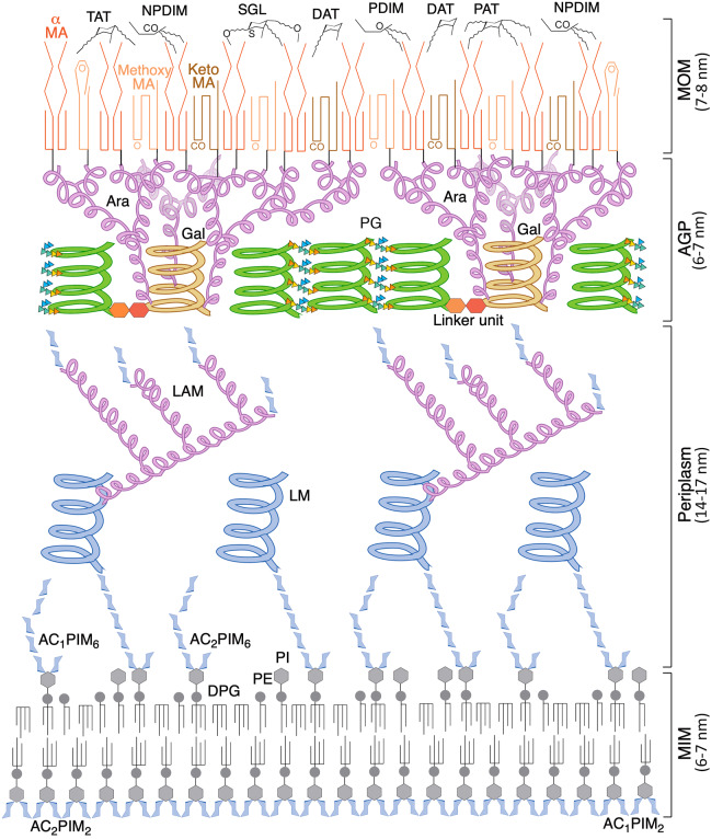
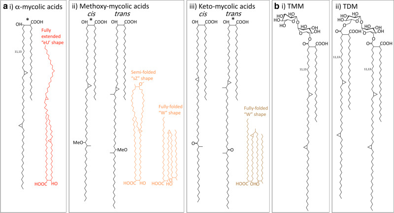
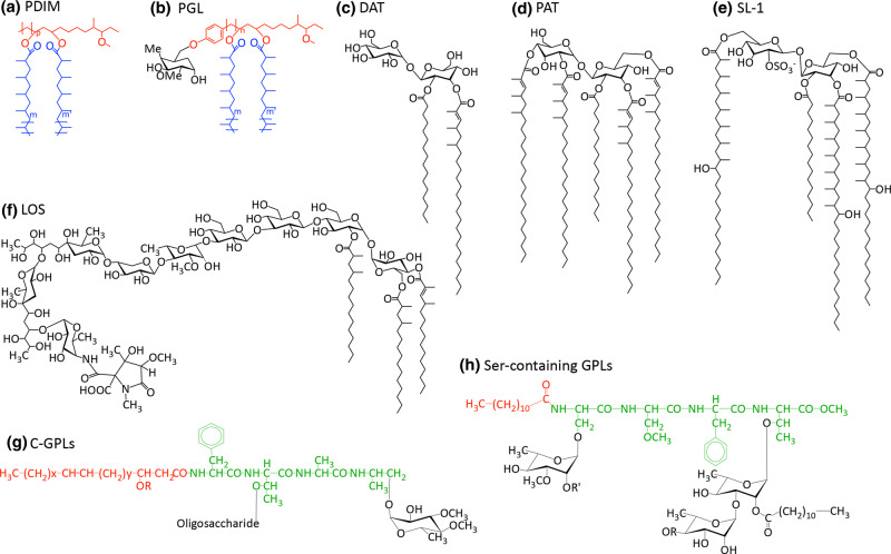
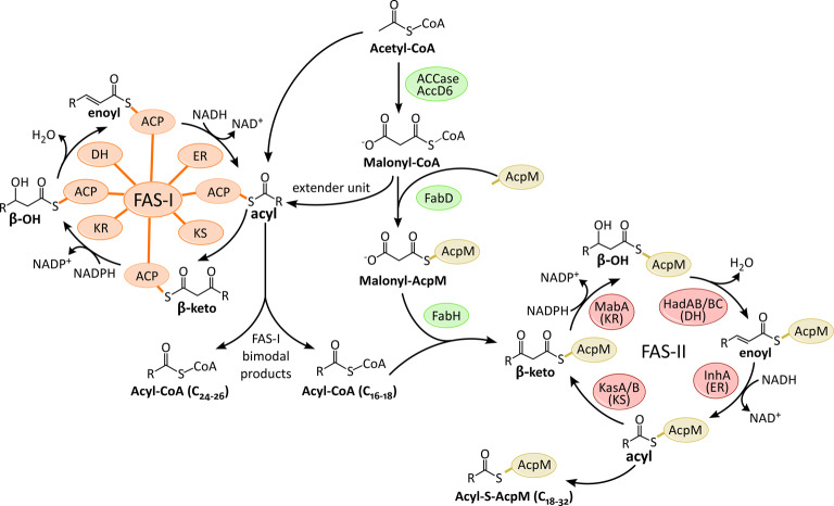
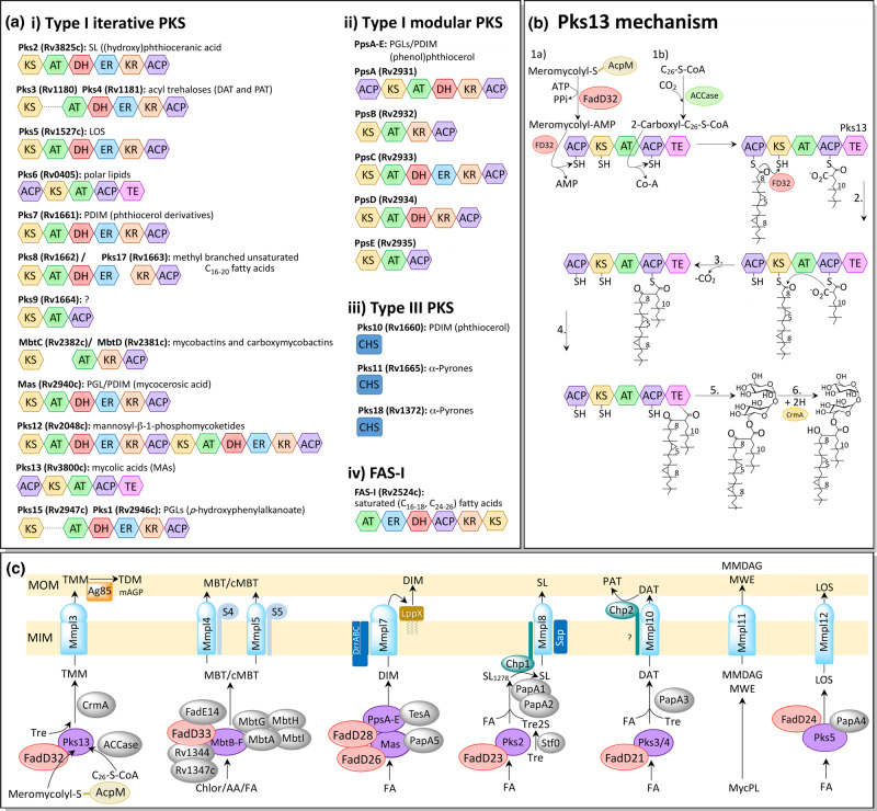
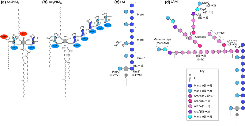
Tuberculosis, caused by the pathogenic bacterium Mycobacterium tuberculosis (Mtb), is the leading cause of death from an infectious disease, with a mortality rate of over a million people per year. This pathogen's remarkable resilience and infectivity is largely due to its unique waxy cell envelope, 40% of which comprises complex lipids. Therefore, an understanding of the structure and function of the cell wall lipids is of huge indirect clinical significance. This review provides a synopsis of the cell envelope and the major lipids contained within, including structure, biosynthesis and roles in pathogenesis.

Keywords: biosynthesis; cell wall; lipids; mycobacterium tuberculosis; mycolic acids.


Conflict of interest statement

The authors declare that there are no competing interests associated with the manuscript.

Figures

Figure 1.
Figure 1.. Structure of the cell envelope of Mtb.
In the MIM (mycobacterial inner membrane), the inner leaflet mostly consists of AC1/2PIM2 (tri-/tetra-acylated phosphatidyl-myo-inositol-dimannoside). The outer leaflet also contains PIMs, in the form of AC1/2PIM6 (tri-/tetra-acylated phosphatidyl-myo-inositol-hexamannoside), as well as the more conventional phospholipids of the inner membrane, DPG (diphosphatidylglycerol), PE (phosphatidylethanolamine) and PI (phosphatidylinositol). In the periplasm, LM (lipomannan) and LAM (lipoarabinomannan) extend outwards, from lipid anchors in the MIM, though additional anchorage in the outer membrane has been demonstrated, which is contentious; the mannose core helix and individual sugar molecules of the PIMs and mannose caps are coloured blue and the branched arabinan domain is pink. The glycan backbone of the PG (peptidoglycan), green, is represented by helices positioned perpendicular to the membrane, according to the scaffold model [281, 282]. Peptide cross-links between the glycan helices are shown as coloured triangles (orange = l-alanine, yellow = d-isoglutamine, green = meso-diaminopimelate and blue = d-alanine). The PG is connected to the Gal (galactan) domain, yellow, of the arabinogalactan (AG) via a linker unit. The branched Ara (arabinan) domain of the AG, pink, is esterified with MA (mycolic acids), comprising the inner leaflet of the MOM (mycobacterial outer membrane); the typical conformation adopted by each class of MA is represented. The outer leaflet of the MOM is characterised by DAT, TAT and PAT (di-/tri- and pentaacyl trehalose); PDIM and NPDIM (dimycocerosates of phthiocerols and phthiodiolones); and SGL (sulphated trehalose glycolipids) [23,283]. The diagram is roughly to scale using dimensions obtained from cryo-electron microscopy [13].
Figure 2.
Figure 2.. Structure of the major mycolic acid (MA) components from Mtb.
(a) MA structures and the typical conformations adopted, (i) α-MAs, (ii) Methoxy-MAs and (iii) Keto-MAs. The main components are denoted with a *. (b) (i) TMM (trehalose monomycolates) and (ii) TDM (trehalose dimycolates) [284].
Figure 3.
Figure 3.. Free lipid structures.
(a) PDIM (Phthiocerol dimycocerosate) and (b) PGL (phenolic glycolipids, general structure): black = saccharide; red = phenolphthiocerol/phthiocerol; blue = multimethyl-branched fatty acids (mycocerosic acids) [285], (c) DAT (diacyl trehaloses) [286], (d) PAT (pentaacyl trehaloses) [286], (e) Sulfolipid-1, SL-1 (sulphated trehalose glycolipid, SGL) [286] (f) Lipooligosaccharides (LOS) from M. marinum [142], (g) C-GPLs (general structure) [103] and (h) Ser-containing GPL from M. xenopi: red = lipid; green = peptide; black = saccharide [106].
Figure 4.
Figure 4.. Biosynthesis of Fatty acids.
For example in the production of mycolic acids (MAs). The FAS-I domains/FAS-II enzymes: KS (β-ketoacyl synthase), KR (β-keto reductase), DH (dehydratase), ER (enoyl reductase) and ACP (acyl carrier protein) [287,288].
Figure 5.
Figure 5.. Polyketide synthase (PKS)/FAS-I domains and synthesis pathways.
(a) Domains. The PKS domains are: AT (acyl transferase; green), ER (enoyl reductase; blue), DH (dehydratase; red), ACP (acyl carrier protein; purple), KR (β-keto reductase; brown), KS (β-ketoacyl synthase; yellow), TE (thioesterase; pink) and CHS (chalcone synthase; dark blue). Other abbreviations: SL = sulfolipids, DAT = diacyl trehaloses, PAT = pentaacyl trehaloses, LOS = lipooligosaccharides, PGL = phenolic glycolipids, PDIM = phthiocerol dimycocerosate [289–291]. (b) Suggested mechanism of Claisen-type condensation by Pks13. 1.a FadD32 (FD32) activates the meromycolyl-S-AcpM with AMP and loads it on the N-terminal ACP (acyl carrier protein) domain of Pks13. 1.b The acyl-CoA (C26) produced by FAS-I is carboxylated by ACCase and loaded onto the C-terminal ACP domain of Pks13 by the AT (acyltransferase) domain. 2. The meromycolyl chain is transferred, in the presence of FD32, to the KS (ketosynthase) domain of PKS13. 3. The Claisen-type condensation reaction: the acidic α-carbon of the carboxyl-acyl group attacks the carbonyl group of the meromycolyl chain, producing 3-oxo-C78-α-mycolate, releasing CO2 and the meromycolyl chain from the KS domain. 4. The TE (thioesterase) domain cleaves the product from the ACP domain, possibly forming a transient covalent bond via an active site serine. 5. The TE domain transfers the product onto a trehalose unit. 6. The keto group is reduced by CmrA, producing the mature trehalose monomycolate (TMM) [138]. (c) MmpL transport of complex lipids across the cytoplasmic membrane in Mtb. PKS (polyketide synthase) proteins are purple, FadD are red and MmpL/S blue. S4 and S5 are MmpS4 and MmpS5, respectively; MmpL4 and 5 are involved in iron scavenging, exporting two siderophores, lipophilic mycobactin (MBT) and hydrophilic carboxymycobactin (cMBT). MmpL11 exports monomero-mycolyl diacylglycerol (MMDAG) and mycolate wax ester (MWE) from the intermediate mycolyl phospholipid (MycPL). Other abbreviations include FA = fatty acid, Tre = trehalose, mAGP = mycoloylarabinogalactan-peptidoglycan [292–294].
Figure 6.
Figure 6.. Structure of phosphatidyl-myo-inositol mannosides, Lipomannan and Lipoarabinomannan.
(a) Ac2PIM2, (b) Ac2PIM6, (c) LM and (d) LAM (and ManLAM). See key for component sugars [23].

Similar articles

Cited by

References

    1. Global Tuberculosis Report (2019) World Health Organization, Geneva, Switzerland
    1. Da Silva A., and Palomino P.E. and C J. (2011) Molecular basis and mechanisms of drug resistance in Mycobacterium tuberculosis: classical and new drugs. J. Antimicrob. Chemother. 66, 1417–1430 10.1093/jac/dkr173 - DOI - PubMed
    1. Asselineau J. and Lederer E. (1950) Structure of the mycolic acids of Mycobacteria. Nature 166, 782–783 10.1038/166782a0 - DOI - PubMed
    1. Ratledge C. (1982) Lipids: cell composition, fatty acid biosynthesis In The Biology of the Mycobacteria (Ratledge C. and Stanford J., eds), pp. 53–94, Academic Press, London, U.K
    1. Minnikin D.E. (1982) Lipids: complex lipids, their chemistry, biosynthesis and role In The Biology of Mycobacteria (Ratledge C. and Stanford J., eds), pp. 95–184, Academic Press, London, U.K

Publication types

MeSH terms